Let's get back to the basics. A #dermtwitter #tweetorial on:
THE PRIMARY LESION!
My plan is to make a #Derm101 series on #morphology and the #skin exam, so this will be the first in that series of #medthreads.
#MedEd #FOAMEd #medtwitter #medstudenttwitter pc:@dermnetnz
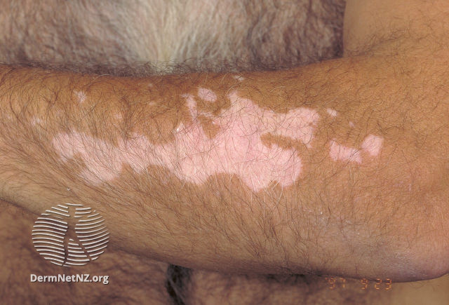
Why are #dermatologists so obsessed with description?
Well, for us, morphology is everything. We start with the exam and take the history afterward based on the possible differential we've come up with!
So let's start simple. What was that lesion in the prior tweet?
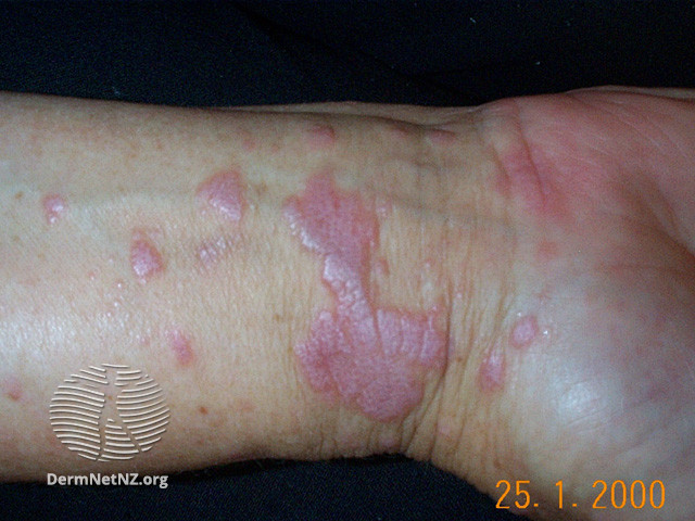
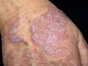
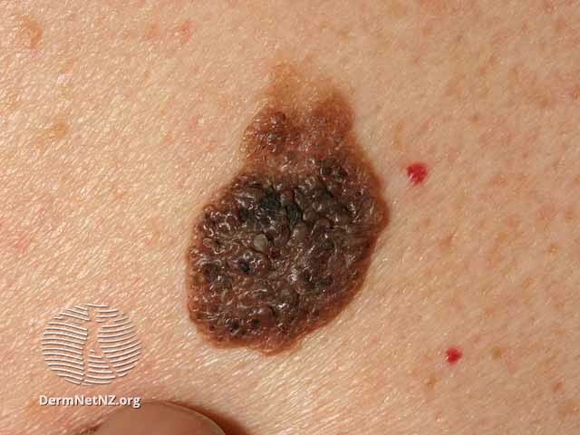
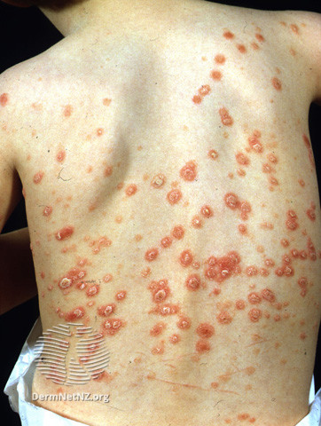
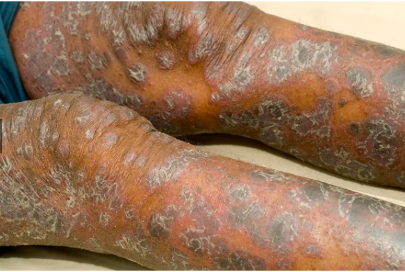
If lesions are raised, they are called PAPULES (<1 cm wide) or PLAQUES (>1 cm wide).
In #1, there's a brown plaque (seborrheic keratosis), & red papules (cherry angiomas).
#2/#3 are examples of papules/plaques of psoriasis in light & dark skin!
pc: healthline.com/health/psorias…
VESICLES AND BULLAE -
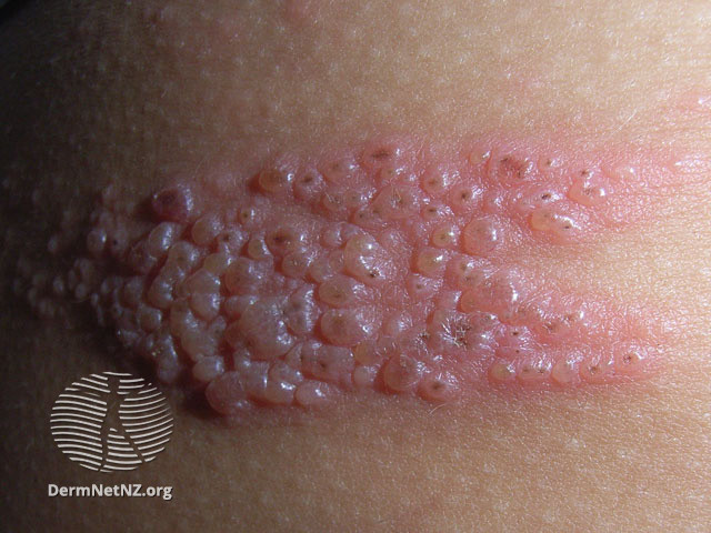
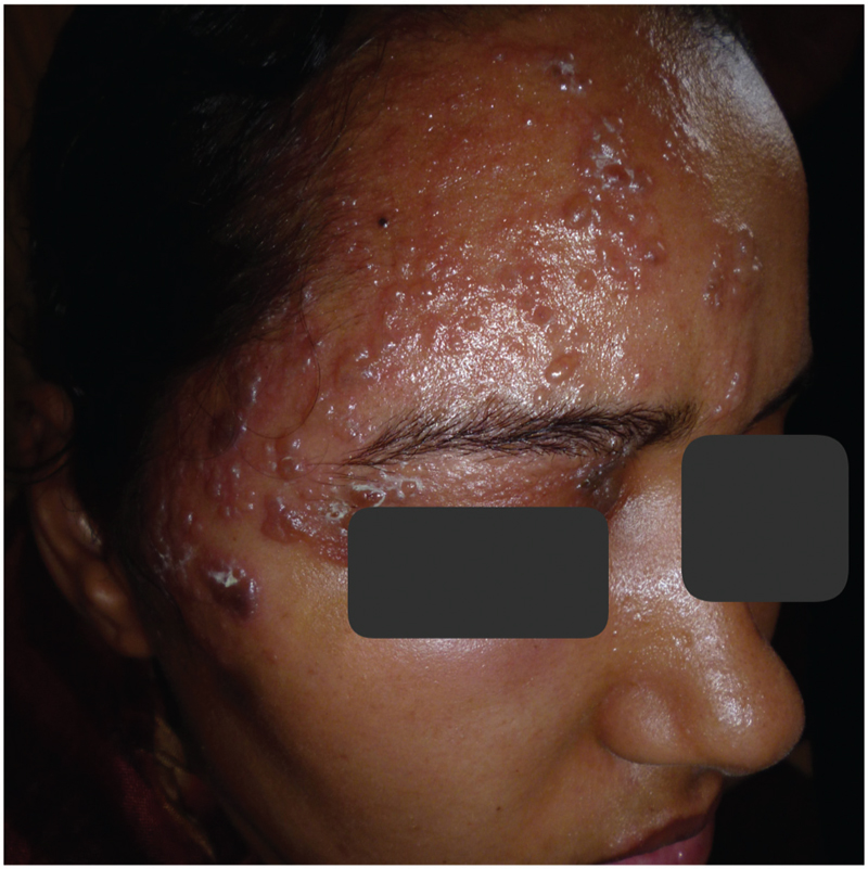
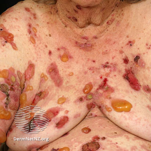
Vesicles are fluid filled lesions <1 cm wide, and Bullae are fluid filled and >1 cm wide!
#1 and #2 are zoster in light and dark skin, where we see vesicles.
#3 is bullous pemphigoid, where we see tense bullae!
pc for #2: ejdv.eg.net/viewimage.asp?…
RECAP: THE PRIMARY LESION
✅Flat: macules (<1 cm), patches (>1 cm)
✅Raised/depressed: papules (<1 cm), plaques (>1 cm)
✅Deeper seated: nodules and tumors
✅Fluid filled: vesicles (<1 cm), bullae (>1 cm)
✅Pustule: pus filled
✅Wheals: hive-like
✅Exam first, history 2nd
Thanks for joining! I know other folks on #dermtwitter have put out similar threads in the past (@HarkerDavid); I couldn't find it, so why not have another!
Also, if you like to learn in video format:
Join me for another #Derm101 #tweetorial soon!


