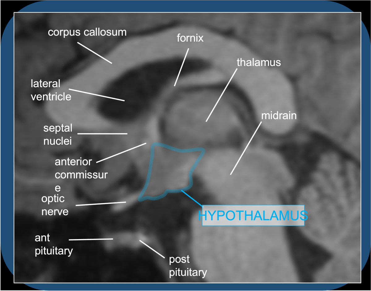
The AC runs across the midline in front of the anterior columns of the fornix, behind the basal forebrain and beneath the anterior limb internal capsule and basal ganglia, surrounded by the bed nucleus of the stria terminalis.
The AC connects areas of the bilateral temporal poles and orbitofrontal cortex. Function is not entirely understood but it is thought to be important in the olfactory pathway and pain sensation, among other things.
Other supratentorial crossing bundles: the corpus callosum, posterior commissure, the hippocampal and habenular commissures. Note: a commissure is a bundle connecting similar regions on both sides; projection fibers and decussations connect different regions at different levels.
The posterior commissure is part of the epithalamus, anterior/inferior to the pineal gland. Important in pupillary light reflex. In MR imaging, a true axial is usually considered as the plane connecting the AC and posterior commissure.
The habenular commissure connects the habenular nuclei of the epithalamus and forms the stalk of the pineal gland. The hippocampal commissure connects the two sides of the body/crus of the fornix.
• • •
Missing some Tweet in this thread? You can try to
force a refresh
Quiz A: Histamine activity in the brain/brainstem contributes to alertness/wakefulness–hence: sleepy effects of benadryl. Histaminergic neurosecretory cell bodies are exclusively in the tuberomammillary nucleus of the TUBER CINEREUM (HT floor), with widespread projections.
2/28
The HT is a mysterious and complex—I get confused sighs from medical students when the topic arises—the small almond-sized morsel is the control center for endocrine/hormone regulation, and homeostasis of food/water consumption, sleep, BP, sex/attachment. The HT is boss!
3/28
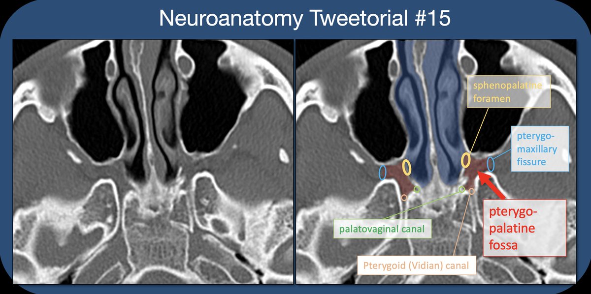
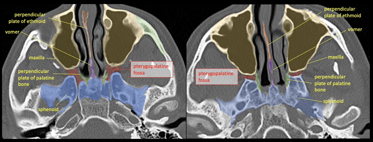
The PTERYGOPALATINE FOSSA (PPF) is a space deep in the face/skull base, bordered anteriorly by the maxilla (max sinus), posteriorly by the pterygoid base of the sphenoid, and medially by the perpendicular plate of the palatine bone. 2/22
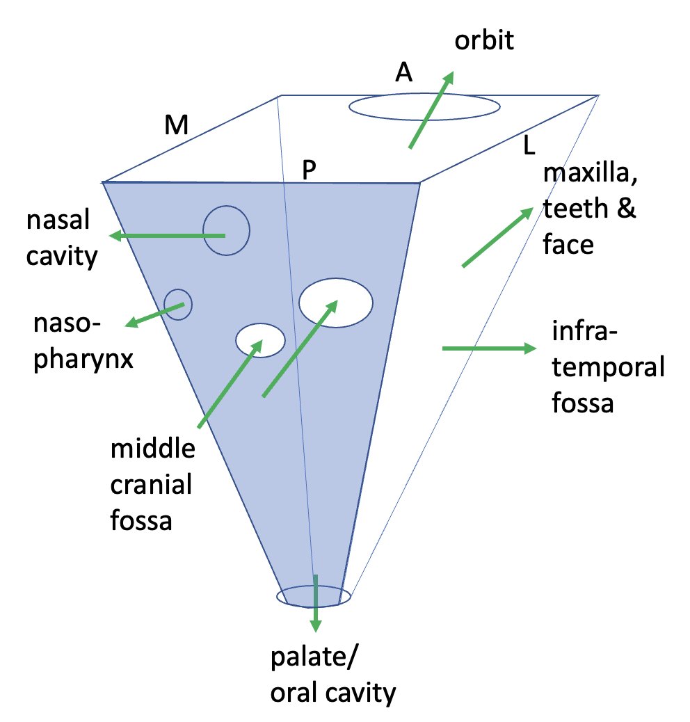
It’s best to think of the PPF as a crossroads/intersection. Think about the roads that lead to and from it, and the cast of characters that pass through.
Some like to simplify/visualize the PPF as a cube or inverted pyramid.
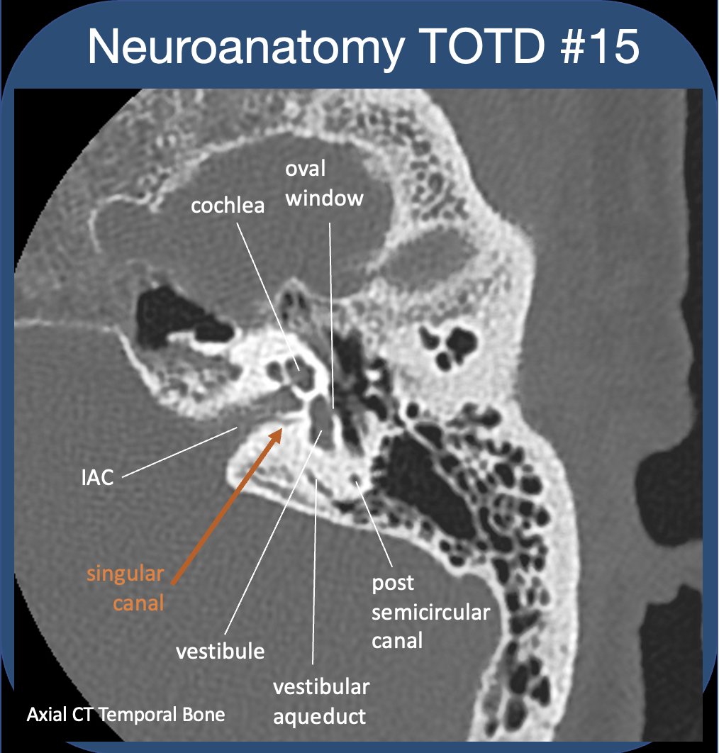
To evaluate the t-bone, best to compartmentalize--external/middle/inner ear (IE). See previous #tweetorial of the ME. The IE is difficult as most structures are obliquely oriented (at different obliquities!)-and can be hard to see on standard views. 2/24
IE communicates with ME via oval&round windows (which allow for transmission&dissipation of sonic vibrations). IE houses sensory organs for hearing/balance/sensing motion. The cochlear&vestibular nerves (CNVIII) transmit signals to the brain via the int auditory canal (IAC). 3/24
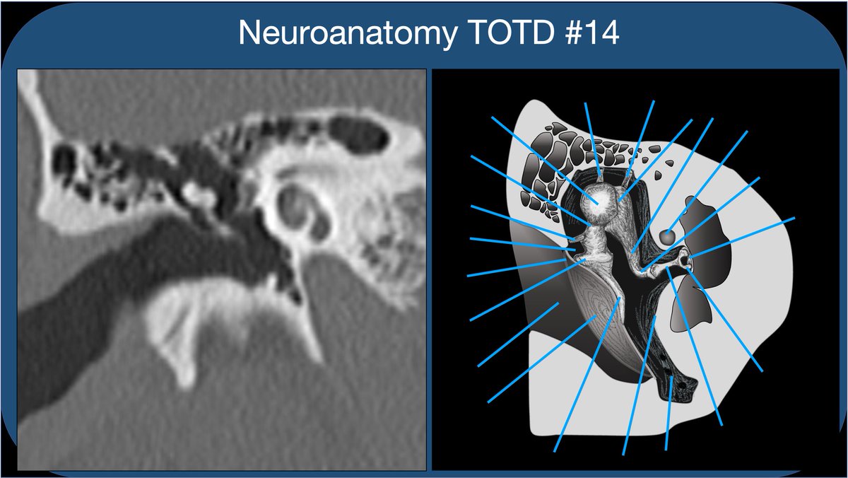
Whether learning t-bone anatomy as a medical student or evaluating a CT of the t-bone as a radres, it’s best to compartmentalize into external, middle, and inner ear. This thread🧵is on the middle ear: Inner ear to follow, some day:)
2/21
The external ear extends from the external meatus to the TM. The TM should be thin and *almost* imperceptible on CT. Thickened and retracted TM suggests prior pathology (usually otitis media) and scarring.
Time for a deep dive into limbic networks. Bear with me—this is a fun subject. I got carried away preparing slides—it’s hard to know when to stop!
2/18
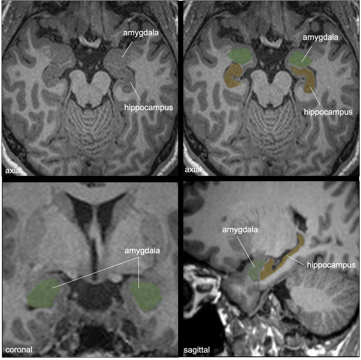
The amygdala is an almond-shaped collection of gray nuclei/subnuclei deep to the uncus and ant to hippocampus in the med temporal lobe. Involved in multiple functions: memory modulation, emotional learning and responses, +important connections w/ the olfactory bulb/cortex.
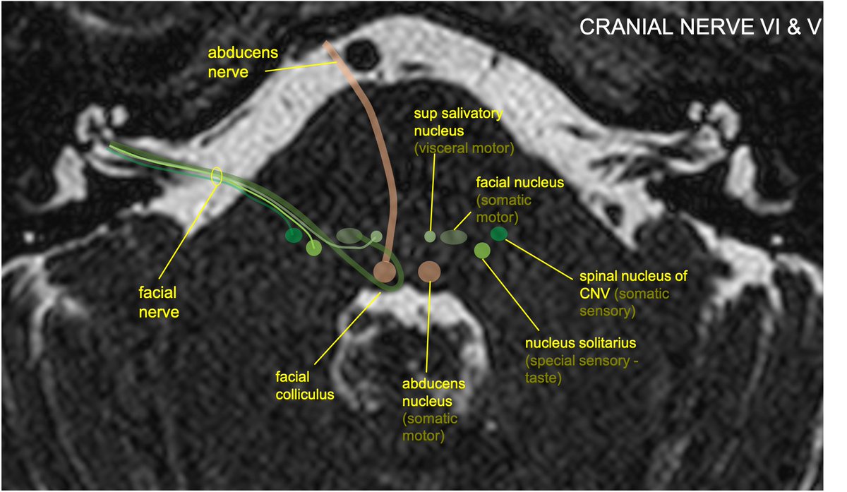
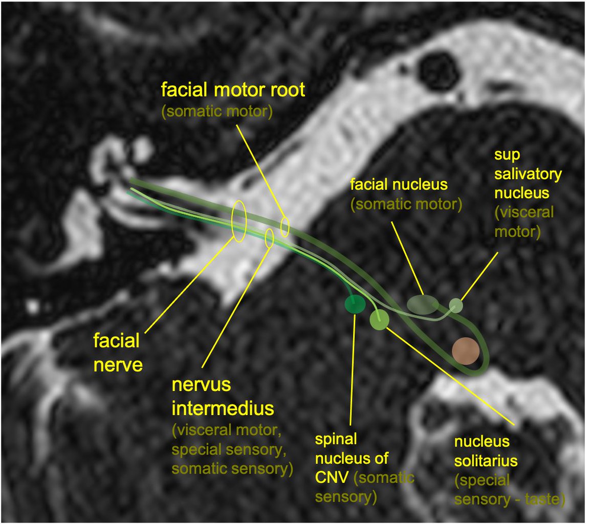
Facial motor nucleus is in the pontine tegmentum; axons loop dorsally around the abducens nucleus, then course anterolaterally, exiting the brainstem at the CPA.
2/11
The nervus intermedius (NI) is composed of preganglionic parasympathetic fibers w/cell bodies in the sup salivatory nucleus, taste fibers w/cell bodies in the nucleus solitarius, and somatic sensory input from EAC/external ear, w/cell bodies in the spinal nucleus of CNV.