Confused about how to perform Lung 🫁 Ultrasound and all those Signs? Don't be!
1⃣Learn a Lung #POCUS Protocol
2⃣Learn all Common Lung US Signs
3⃣Learn all Major Lung US Pathologies
✅New Blog Post!
👉🔗pocus101.com/Lung
#medtweetorial 👇 (1/24)


1⃣Learn a Lung #POCUS Protocol
2⃣Learn all Common Lung US Signs
3⃣Learn all Major Lung US Pathologies
✅New Blog Post!
👉🔗pocus101.com/Lung
#medtweetorial 👇 (1/24)


(2/) The parietal pleura interfaces with the visceral pleura, creating a sliding motion as we breathe.
Alveoli exist in lobules that are subdivided by interlobular septa. These areas can fill with fluid due to consolidation or pulmonary edema.
👉🔗pocus101.com/Lung
Alveoli exist in lobules that are subdivided by interlobular septa. These areas can fill with fluid due to consolidation or pulmonary edema.
👉🔗pocus101.com/Lung

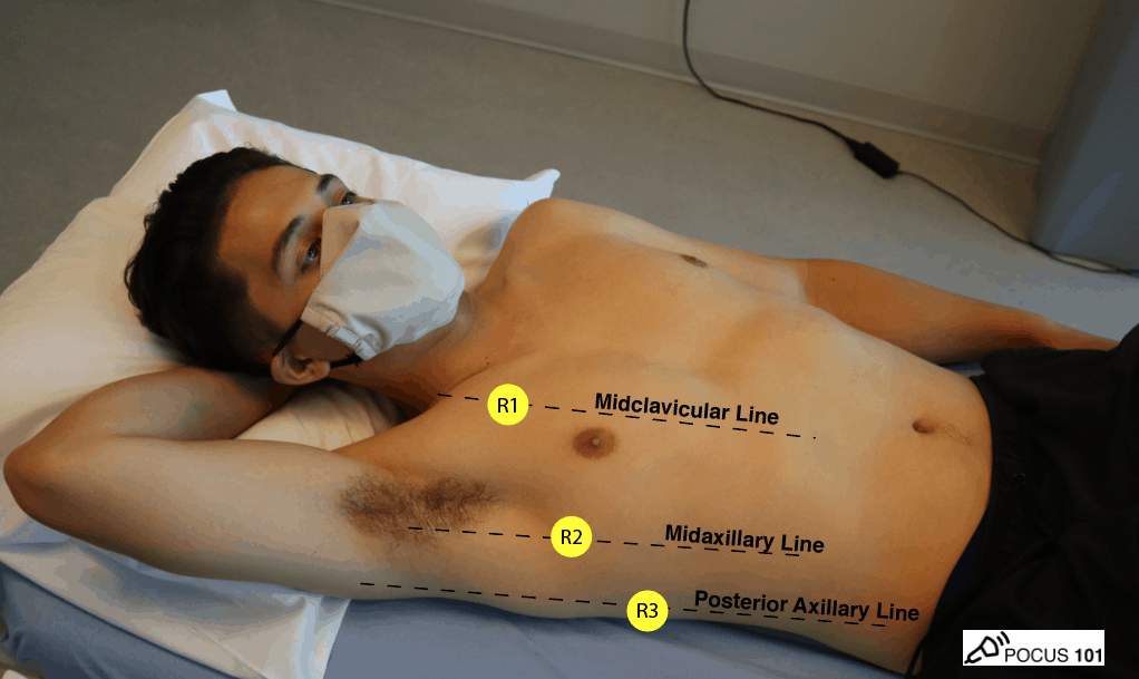
(4/) At our shop, we use a 6-point lung ultrasound protocol for general lung ultrasound. We expand this to 12 points if needed.
👉🔗pocus101.com/Lung


👉🔗pocus101.com/Lung


(6/) Identify Lung Sliding using B-Mode
👉🔗pocus101.com/Lung
👉🔗pocus101.com/Lung
(7/) Evaluate for lung sliding using M-Mode by identifying the Seashore sign for the "sky, ocean, and beach."
👉🔗pocus101.com/Lung
👉🔗pocus101.com/Lung

(8/) Identify A-lines by placing your probe perpendicular to the pleura.
👉🔗pocus101.com/Lung
👉🔗pocus101.com/Lung
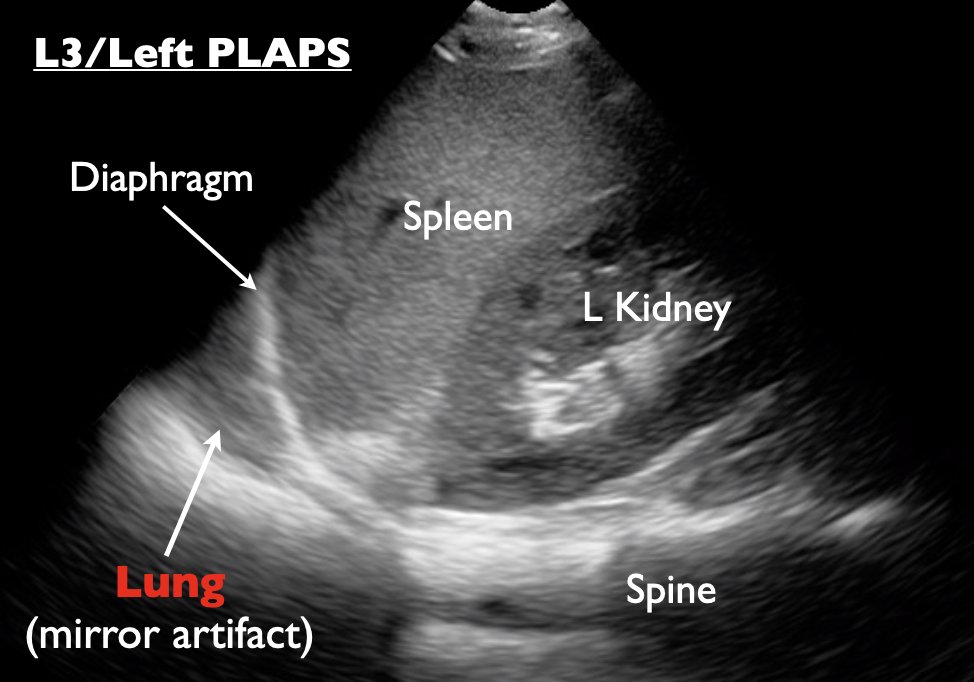
(10/) The curtain sign is seen in healthy and aerated lungs at PLAPS point. An aerated lung is like a “curtain” because as it fills with air, it looks like a curtain sweeping down and over the other organs, momentarily obscuring them from view.
👉🔗pocus101.com/Lung
👉🔗pocus101.com/Lung
(12/) Absent Lung Sliding with B-mode does not just mean PTX. It can ALSO be from:
pleurodesis
inflammation (ARDS)
severe COPD
Fibrotic lungs
Mainstem intubation
👉🔗pocus101.com/Lung
pleurodesis
inflammation (ARDS)
severe COPD
Fibrotic lungs
Mainstem intubation
👉🔗pocus101.com/Lung
(13/) Absent lung sliding with M-Mode (the Barcode or Stratosphere signs).
👉🔗pocus101.com/Lung
👉🔗pocus101.com/Lung
(14/) To rule in PTX look for the Lung Point Sign
👉🔗pocus101.com/Lung
👉🔗pocus101.com/Lung
(15/) For Pneumonias look for Dynamic Airbronchgrams within your consolidation.
👉🔗pocus101.com/Lung
👉🔗pocus101.com/Lung
(16/) Finding it difficult to keep track of all the signs for pleural effusion? Don't worry, we got you covered:
👉🔗pocus101.com/Lung
Spine Sign
Jellyfish sign
Quad Sign
Sinusoid Sign
Plankton Sign
Hematocrit Sign
Loculated Pleural Effusions
👉🔗pocus101.com/Lung
Spine Sign
Jellyfish sign
Quad Sign
Sinusoid Sign
Plankton Sign
Hematocrit Sign
Loculated Pleural Effusions
(17/) Spine Sign with spine extending past the diaphragm superiorly means there is a consolidation or pleural effusion present.
👉🔗pocus101.com/Lung
👉🔗pocus101.com/Lung
(18/) The “Jellyfish Sign” occurs when a consolidated lung is seen floating in the pleural effusion.
👉🔗pocus101.com/Lung
👉🔗pocus101.com/Lung
(19/) You can turn on M-mode to look for the Sinusoid sign (looks like a sine wave). It is caused by the parietal and visceral pleura moving closer and further apart while the patient breathes.
👉🔗pocus101.com/Lung
👉🔗pocus101.com/Lung

(20/) A pleural effusion has an anechoic appearance often delineated by the pleural line, the rib shadows, and the lung line, called the “Quad Sign.”
👉🔗pocus101.com/Lung

👉🔗pocus101.com/Lung


(21/) The plankton sign shows an effusion with swirling, hyperechoic debris.
👉🔗pocus101.com/Lung
👉🔗pocus101.com/Lung
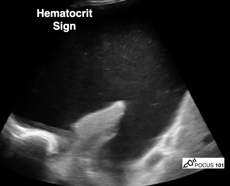
(22/) The hematocrit sign refers to the echogenic layering of material in a pleural effusion. This can be due to exudative effusions or a hemothorax.
👉🔗pocus101.com/Lung
👉🔗pocus101.com/Lung
(23/) Here is an example of loculated pleural effusion
👉🔗pocus101.com/Lung
👉🔗pocus101.com/Lung
(24/) Want more details on all of the major Lung Ultrasound Pathology Profiles? Check out the FULL blog post that goes over everything!
👉🔗pocus101.com/Lung
👉🔗pocus101.com/Lung

Don't forget to check out the #POCUS clinical review book for a ton of Lung Ultrasound Cases!
Click here for a free preview of the book and an exclusive 20% discount: pocus101.com/RevBook
Click here for a free preview of the book and an exclusive 20% discount: pocus101.com/RevBook

• • •
Missing some Tweet in this thread? You can try to
force a refresh


