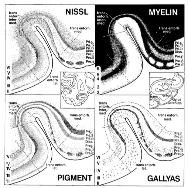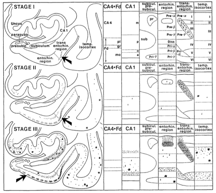
Hello and happy #SubfieldWednesday! Today we are going to get a bit more familiar with how the hippocampal subfields differ in their composition of different cell types, cell sizes, and layer thickness. 🍤🔬
#SubfieldWednesday (1/n)
#SubfieldWednesday (1/n)
Here are some images taken from five different hippocampal subfields (CA1, CA2, CA3, dentate gyrus, and subiculum). Can you tell which number corresponds to which subfield? 🤔
#SubfieldWednesday (2/n)
#SubfieldWednesday (2/n)

Because a Nissl stain was applied to these slices, the cell bodies appear dark purple. This allows neuroanatomists to characterize the size, shape, and relative spacing of the cells.
#SubfieldWednesday (3/n)
#SubfieldWednesday (3/n)
Here are some hints to help you identify them. Let's start with the most obvious: dentate gyrus (DG)
DG is easily identifiable because of the densely packed layer of granule cells. These cells are smaller & rounder in shape compared to pyramidal cells
#SubfieldWednesday (4/n)
DG is easily identifiable because of the densely packed layer of granule cells. These cells are smaller & rounder in shape compared to pyramidal cells
#SubfieldWednesday (4/n)
Next we will describe how CA1, CA2, CA3, & subiculum differ. 🧐
Pyramidal cell bodies (somata) within CA1 are triangular in shape, smaller than those of CA2 and CA3, and more scattered
#SubfieldWednesday (5/n)
Pyramidal cell bodies (somata) within CA1 are triangular in shape, smaller than those of CA2 and CA3, and more scattered
#SubfieldWednesday (5/n)
The CA2 region has the most compact and narrow pyramidal cell layer of the hippocampus. Its cells are about as large and darkly stained as those in CA3, but there is far less space between the cell bodies.
#SubfieldWednesday (6/n)
#SubfieldWednesday (6/n)
The CA3 pyramidal cells are the largest of the hippocampus and stain darkly in Nissl stains. While the layer is approximately ten cells thick, it nonetheless has a relatively homogeneous appearance.
#SubfieldWednesday (7/n)
#SubfieldWednesday (7/n)
Finally, the pyramidal cell layer of the subiculum is 30 or more cells thick, and can be divided into at least two sublaminae (sub-layers). Relative to CA1, the subicular pyramidal cells tend to be somewhat larger and more widely spaced.
#SubfieldWednesday (8/n)
#SubfieldWednesday (8/n)
After reading the thread, do you think you can identify all five subfields correctly? Test your knowledge in the quiz below.
#SubfieldWednesday (9/n)
#SubfieldWednesday (9/n)

One more thing! A big thank you to Ricardo Insausti and Paul Yushkevich for providing the histological sections used in this thread!
@threadreaderapp unroll
• • •
Missing some Tweet in this thread? You can try to
force a refresh


