Known distal UVJ stone. Has new rt flank pain. Mild hydro but what else? #POCUS #IMPOCUS #FOAMed #FOAMus #MedEd #MedTwitter @ACEP_EUS @SeeWithSound @PhilipsPOCUS @IMPOCUSFocus @medpedshosp @NephroP @jminardi21 @HeyDrNik @TomJelic @cianmcdermott @MH_EMultrasound @thepocusatlas
Area of increased echogenicity of parenchyma seen in upper pole. @Manoj_Wickram @tomkehrl @MH_EMultrasound @MetroHealth_EM @Casscln @ria_dancel @cm_lopresti @DRsonosRD
Also has trace perinephric fluid seen (arrow)
Doppler showed decreased perfusion in the area of increased echogenicity. Diagnosis?
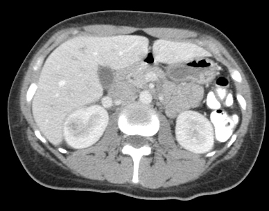
Diagnosis: acute focal nephritis. Also called focal lobar nephronia by rads. This is on spectrum somewhere between acute pyelonephritis and renal abscess. CT shown. Contrast image (not shown) demonstrated area of poorly perfused parenchyma without a cortical rim sign.
Since had know obstructing stone, the trace perinephric fluid could be a urinoma vs secondary to infection.
Primary role of u/s in potential renal infection is to evaluate for obstruction (known in this pt) or higher grade infection such as renal abscess or perinephric abscess. In most cases of uncomplicated pyelo the ultrasound will be normal.
Other findings include urothelial thickening. Reliable finding in correct clinical context. However, can be seen with stones, stents, and cancer. Here is urothelial thickening in another patient with higher grade pyelonephritis.
Video clip of patient with higher grade pyelonephritis with urothelial thickening.
Pyonephrosis can also be seen in renal infections. If echoes seen within the collecting system differential would be blood vs. pus vs. mass. Clinical correlation important. Pyonephrosis in patient with distal obstructing stone.
Pyonephrosis seen layered in dilated collecting system on TRV image in patient with proximal stone which is also seen.
Renal abscess: Circular area with internal echoes and hyperechoic rim since in lower pole of left kidney.
Doppler image of renal abscess. No internal flow. Since abscesses appear as complex cystic masses could be confused with tumors. You should not see Doppler flow within a renal abscess (same as with soft tissue or other abscesses). Further imaging needed if diagnosis in doubt.
Another patient with renal abscess.
Xanthogranulomatous pyelonephritis-chronic process. Proteus mirabilis and E.coli common organisms. Commonly associated with staghorn calculi as seen in this patient.
Emphysematous pyelonephritis is a serious infection due to gas forming organisms where air will be identified within the parenchyma (see video) as compared to emphysematous pyelitis where air is only seen in collecting system.
Air within bladder on sag clip. This could be the result of an emphysematous cystitis or pyelitis but could also be iatrogenic due to recent Foley catheter placement or other instrumentation. Also note in this patient a high grade infection, the presence of layered echoes.

• • •

Missing some Tweet in this thread? You can try to force a refresh
 

Keep Current with Robert Jones

Robert Jones Profile picture

Stay in touch and get notified when new unrolls are available from this author!

Read all threads

This Thread may be Removed Anytime!

PDF

Twitter may remove this content at anytime! Save it as PDF for later use!

Try unrolling a thread yourself!

how to unroll video
  1. Follow @ThreadReaderApp to mention us!

  2. From a Twitter thread mention us with a keyword "unroll"
@threadreaderapp unroll

Practice here first or read more on our help page!

More from @RJonesSonoEM

3 Jun 20
Differentiating portal venous gas from pneumobilia on ultrasound. See thread. #POCUS #FOAMus #FOAMed #IMPOCUS #MedEd #MedTwitter #ultrasound @ACEP_EUS @USIGcleveland @MetroHealth_EM @aedrake65
Case 1. Liver TRV sweep. Which is present? Answer poll.
Case 1. Which is present?
Read 9 tweets
19 May 20
Pt-Bilat LE pain/swelling 5 days.Prior hx of unprovoked DVT/PE years ago but off meds due to insurance. See thread. POCUS DVT #POCUS #IMPOCUS #FOAMed #FOAMus #MedEd #FOAMcc @ACEP_EUS @medpedshosp @cianmcdermott @jminardi21 @grepmeded @siddharth_dugar @msiuba @NephroP @SonoStache
What POCUS DVT protocol to use? 2-point or 3-point or extended? With or without Doppler? Meta-analysis showed good performance of 2 and 3 point POCUS but need to understand POCUS compression limitations if only compression used: pubmed.ncbi.nlm.nih.gov/31145304/?from…
Adhikari and colleague article showed 6.3% false neg rate due to prox isolated DVT with 2-point compression. Limitations of POCUS prox compression DVT exam go beyond just missing isolated/focal DVT. pubmed.ncbi.nlm.nih.gov/25465473/
Read 9 tweets
12 May 20
Patient with RUQ abdominal pain. Difficulty finding GB. Pt denies history of prior abdominal surgery and no scars seen. See thread. #POCUS #IMPOCUS #FOAMed #FOAMus #MedEd #MedStudentTwitter @medpedshosp @NephroP @grepmeded @cianmcdermott @ACEP_EUS @PhilipsPOCUS
GB Sag and TRV sweeps. GB not seen. No surgical clips seen. Contracted GB, agenesis of GB, ectopic location of GB, prior removal (and patient forgot--it does happen)?
On Sag image, don't confuse bowel contents with GB filled with sludge
Read 6 tweets
24 Mar 20
Short image tweetorial on thoracic ultrasound findings in pneumonia. #POCUS #IMPOCUS #FOAMed #FOAMus #Medtwitter #Meded #COVID19 @medpedshosp @cianmcdermott @msiuba @siddharth_dugar @grepmeded @NephroP @emresidents
Normal lung ultrasound. A-profile with thin pleural line with lung sliding. Linear transducer used.
Irregular pleural line with patchy B-lines. Seen in viral (including COVID19) and bacterial pneumonia. This patient had mycoplasma pneumonia. Linear probe used.
Read 11 tweets

Did Thread Reader help you today?

Support us! We are indie developers!


This site is made by just two indie developers on a laptop doing marketing, support and development! Read more about the story.

Become a Premium Member ($3/month or $30/year) and get exclusive features!

Become Premium

Too expensive? Make a small donation by buying us coffee ($5) or help with server cost ($10)

Donate via Paypal Become our Patreon

Thank you for your support!

Follow Us on Twitter!