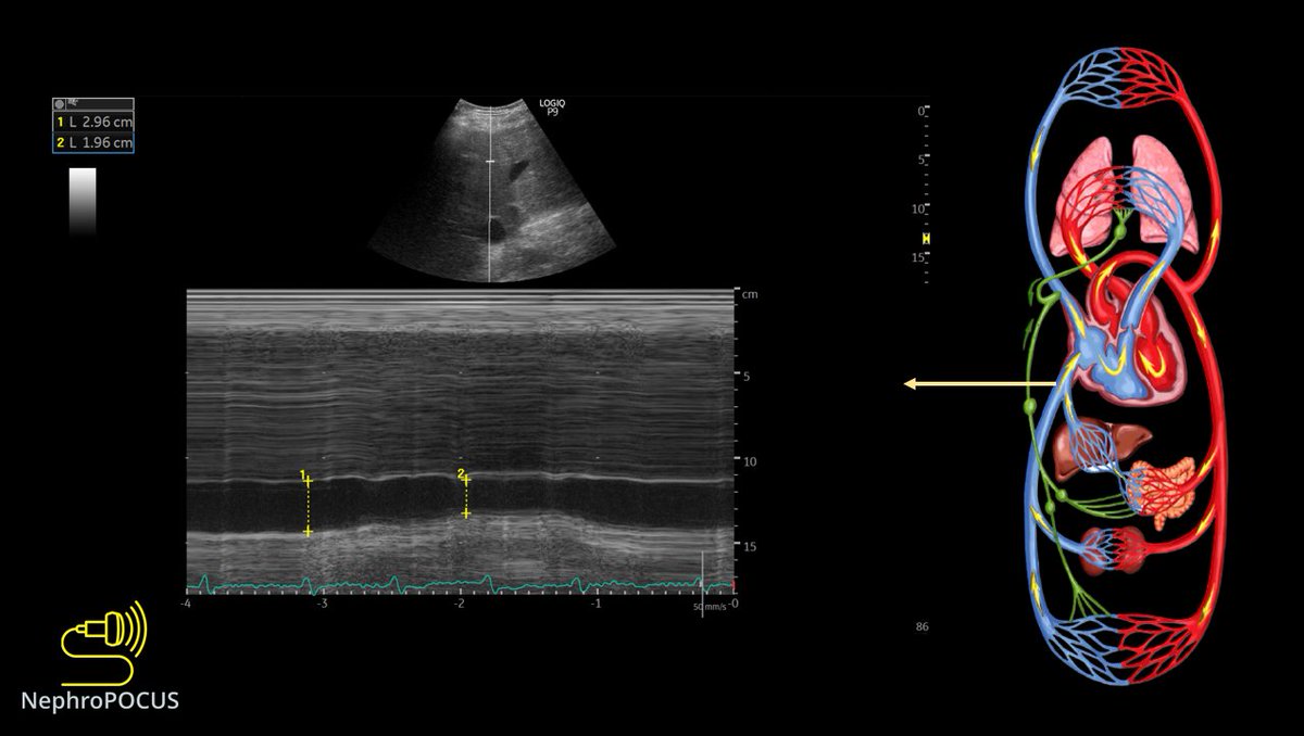
#POCUS quiz of the day:
Easy one. Identify 1, 2 and 3.
POLL in thread πŸ‘‡ #IMPOCUS #MedEd
πŸ‘†#POCUS
PE = pleural effusion
PER = pericardial effusion
Ao = aorta
IVC = inferior vena cava
Rt = right
Lt = left
Correct answer: option 2 -
1. left pleural effusion (note the appearance of collapsed lung; also u can see rib shadows/posterior chest wall = lung area)
2. Right pleural effusion (remember the Boomerang sign on subxiphoid view?)
3. IVC

β€’ β€’ β€’

Missing some Tweet in this thread? You can try to force a refresh
γ€€

Keep Current with NephroPOCUS

NephroPOCUS Profile picture

Stay in touch and get notified when new unrolls are available from this author!

Read all threads

This Thread may be Removed Anytime!

PDF

Twitter may remove this content at anytime! Save it as PDF for later use!

Try unrolling a thread yourself!

how to unroll video
  1. Follow @ThreadReaderApp to mention us!

  2. From a Twitter thread mention us with a keyword "unroll"
@threadreaderapp unroll

Practice here first or read more on our help page!

More from @NephroP

24 Apr
#POCUS #echofirst #MedEd case of the day.
Context: alcoholic liver cirrhosis with suspected #hepatorenal syndrome
Purpose: think about the underlying pathophysiology & seek expert input
1/ PLAX, PSAX, Apical views of the heart. Heart rate ~110-116 bpm
#VExUS images in 🧡
2/ overall, looks hyperdynamic. LA looks little enlarged on PLAX, visual TAPSE high on apical.
In cirrhosis, hyperdynamic circulation is expected because of splanchnic vasodilatation and 'relative' arterial under filling.
LV EF #POCUS obtained using @kosmosplatform πŸ‘‡ Image
3/ Lets calculate stroke volume using LVOT VTI #POCUS
It looks supra-normal Image
Read 20 tweets
23 Apr
OK #VExUS #POCUS enthusiasts, time for another case discussion.
Somebody asked if I ever recommend IV fluid in a patient with #VExUS 3.
Here is one example where I did.
1/ First, let's see the #physicalexam (#IMPOCUS) findings, then will tell about the case. #MedEd #Nephrology Image
2/ So, hepatic shows D-only patternπŸ‘†
If we are doing #VExUS, IVC must be big. Here is the M-mode #POCUS πŸ‘‡ Image
3/ Renal parenchymal vein #VExUS #POCUS
It also demonstrates D-only pattern πŸ‘‡ Image
Read 15 tweets
9 Apr
Time for a #POCUS #tweetorial on optimization of Doppler. Very important for #VExUS enthusiasts. #MedEd
1/ Unlike greyscale imaging which depends on amplitude of the returned signal, Doppler depends on frequency information. This graphic explains why perpendicular angle is bad.
2/ other way of saying this, in relevance to color Doppler #POCUS
RBC moving away from the probe = Fr<Ft = negative Doppler shift = Blue color
RBC moving towards = Fr>Ft = positive Doppler shift = Red color
Rest of the images/videos from this excellent paper: pubs.rsna.org/doi/10.1148/rg…
3/ Anatomy (components) of a spectral Doppler waveform (carotid shown)πŸ‘‡
Above baseline is like red on color (towards probe), below = blue. As 0 degree angle is not always possible, <60 is considered OK.
Read 19 tweets
8 Apr
@khaycock2 @ArgaizR @katiewiskar @ThinkingCC
Any comments on this portal vein pulsatility obtained from a pt with cirrhosis? (Why prox is more pulsatile?)
No cardiac issue that I know of; was reviewing rad-performed images πŸ€”
Splenic seems to be fine, looks more like that of distal portal.
Hepatics
Read 4 tweets
6 Apr
1/ Thought of doing a quick #tweetorial on image acquisition for #POCUS friends starting to do #VExUS
It's kind of "how I do it" guide and not necessarily optimized for research.
1st: Look at the IVC in both long and short axes
If big, do further scans πŸ‘‡ #MedEd #FOAMed
2/ Lateral approach works best to obtain a straight segment of the portal vein (straight = best Doppler shift) and a nice hepatic vein too.
Place transducer approximately in the anterior axillary line pointing towards sternal notch. Then fan antero-posteriorly.
#POCUS #VExUS
3/ Forgot what is fanning?
Its also called tilting or some people say, "look" in a particular direction from the same spot.
#POCUS
Read 18 tweets
15 Feb
#Nephrology #POCUS case of the day:
What do you think is this anechoic structure adjacent to liver?
See thread πŸ‘‡ for the answer and more images. #MedEd #IMPOCUS
Let's start with a poll before seeing other images: ☝️? #POCUS
The answer is right renal cyst. Note how the kidney appears with fanning the probe. #POCUS
Is rest of the kidney normal? doesn't appear to be...πŸ€”
Read 11 tweets

Did Thread Reader help you today?

Support us! We are indie developers!


This site is made by just two indie developers on a laptop doing marketing, support and development! Read more about the story.

Become a Premium Member ($3/month or $30/year) and get exclusive features!

Become Premium

Too expensive? Make a small donation by buying us coffee ($5) or help with server cost ($10)

Donate via Paypal Become our Patreon

Thank you for your support!

Follow Us on Twitter!