#Nephrology#POCUS case of the day:
What do you think is this anechoic structure adjacent to liver?
See thread π for the answer and more images. #MedEd#IMPOCUS
Let's start with a poll before seeing other images: βοΈ? #POCUS
The answer is right renal cyst. Note how the kidney appears with fanning the probe. #POCUS
Is rest of the kidney normal? doesn't appear to be...π€
More cysts? hydronephrosis?
Remember this #POCUS flashcard? π
We know that hydronephrosis is like a branching tree and cyst is like a ball. We see both here: #POCUS
Another #POCUS image showing more right renal cysts
So this kidney has cysts + hydronephrosis: a tricky combo!
Was the initial large cyst simple or complex?
Remember the definition? - any cyst that is not simple is complex π
Read this quick #POCUS post nephropocus.com/2019/06/20/renβ¦
As the cyst has a septation βοΈ, it is complex.
How does the patient's left kidney look like? Also has multiple cysts; no significant hydronephrosis π
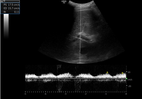
Looks D-only but could this be S-wave? (or a delayed D? Pt has Afib and predisposed to having smaller S but wondering if there is some S-D fusion here)
Renal #POCUS in a #dialysis patient: Looks fine at first glance (thin parenchyma as expected) but fanning anteriorly reveals a large cyst, which would have been missed otherwise. Labeled images and notes in thread π #Nephrology#MedEd#IMPOCUS
Labeled first image #POCUS
Note: peri-renal fat (usually prominent in ESRD patients) can be confused with free fluid.
Labeled second image #POCUS (obtained with phased array probe)
Note: EKG is not needed π
#Nephrology consult for worsening creatinine in the context of diuresis in a patient with #heartfailure
Documented weight 3 kg β¬οΈ than the previous day. No accurate urine output. Pt feels OK (has some unrelated issue).
Question: over diuresis? #POCUS#MedEd#VExUS see thread π
So, there is decreased LV systolic function and obviously diastolic dysfunction. Bi atrial enlargement. No significant pericardial effusion (there was trace on PLAX).
Next step in the hemodynamic circuit #POCUS assessment?
btw repeat weight was similar.
Next: IVC #POCUS assessment to assess right atrial pressure.
Here it is π
Big and plethoric (take it as 15 mmHg in a spontaneously breathing person - we get RVSP of ~41 mmHg based on the above tricuspid gradient)
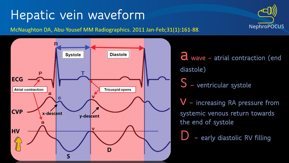
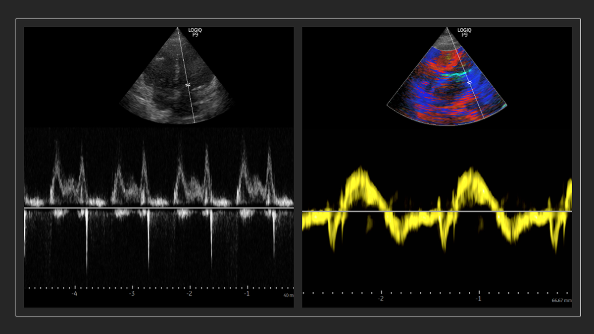
#POCUS Intra-renal vein Doppler waveform looking exactly like that of hepatic vein! #VExUS obtained from a #nephrology clinic patient with pulmonary hypertension. #MedEd
@Thind888@FH_Verbrugge@khaycock2 Dear hemodynamic masters, saw a cirrhotic pt with oliguria (relatively preserved Scr) & pulmonary congestion. Based on these findings, suggested norepinephrine (pressors). Agree? any educational input appreciated.
Basically I interpreted as high output cardiac failure with elevated filling pressures leading to pulmonary congestion.
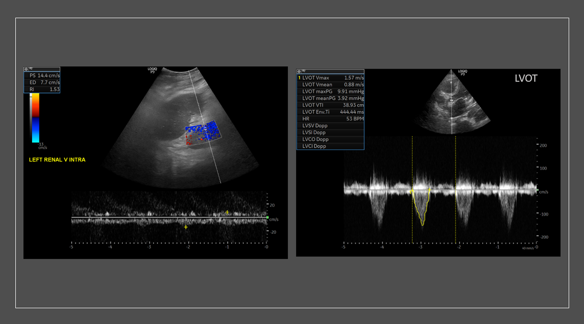
This tracing I believe is right intra-renal 'arterial' with diminished diastolic flow. Correct me if I'm wrong.