@khaycock2 @ArgaizR @katiewiskar @ThinkingCC
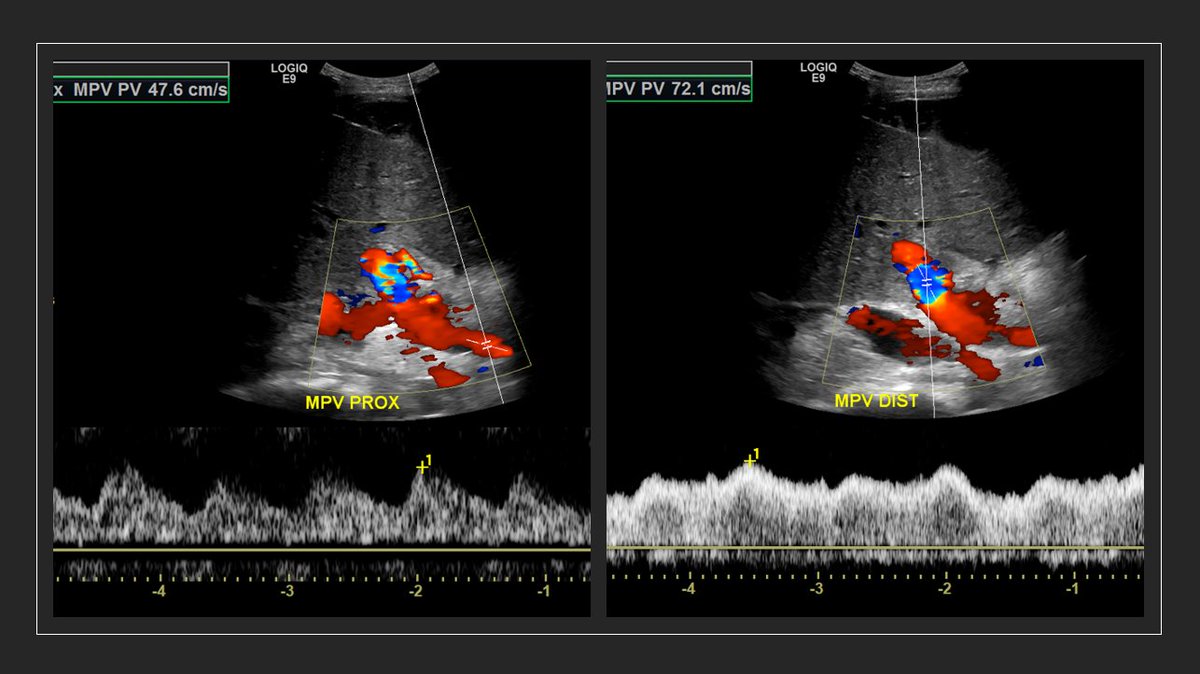
Any comments on this portal vein pulsatility obtained from a pt with cirrhosis? (Why prox is more pulsatile?)
No cardiac issue that I know of; was reviewing rad-performed images 🤔
Splenic seems to be fine, looks more like that of distal portal.
Hepatics
Anything to do with hepatic artery position 🤔

• • •

Missing some Tweet in this thread? You can try to force a refresh
 

Keep Current with NephroPOCUS

NephroPOCUS Profile picture

Stay in touch and get notified when new unrolls are available from this author!

Read all threads

This Thread may be Removed Anytime!

PDF

Twitter may remove this content at anytime! Save it as PDF for later use!

Try unrolling a thread yourself!

how to unroll video
  1. Follow @ThreadReaderApp to mention us!

  2. From a Twitter thread mention us with a keyword "unroll"
@threadreaderapp unroll

Practice here first or read more on our help page!

More from @NephroP

9 Apr
Time for a #POCUS #tweetorial on optimization of Doppler. Very important for #VExUS enthusiasts. #MedEd
1/ Unlike greyscale imaging which depends on amplitude of the returned signal, Doppler depends on frequency information. This graphic explains why perpendicular angle is bad. Image
2/ other way of saying this, in relevance to color Doppler #POCUS
RBC moving away from the probe = Fr<Ft = negative Doppler shift = Blue color
RBC moving towards = Fr>Ft = positive Doppler shift = Red color
Rest of the images/videos from this excellent paper: pubs.rsna.org/doi/10.1148/rg…
3/ Anatomy (components) of a spectral Doppler waveform (carotid shown)👇
Above baseline is like red on color (towards probe), below = blue. As 0 degree angle is not always possible, <60 is considered OK. Image
Read 19 tweets
6 Apr
1/ Thought of doing a quick #tweetorial on image acquisition for #POCUS friends starting to do #VExUS
It's kind of "how I do it" guide and not necessarily optimized for research.
1st: Look at the IVC in both long and short axes
If big, do further scans 👇 #MedEd #FOAMed
2/ Lateral approach works best to obtain a straight segment of the portal vein (straight = best Doppler shift) and a nice hepatic vein too.
Place transducer approximately in the anterior axillary line pointing towards sternal notch. Then fan antero-posteriorly.
#POCUS #VExUS
3/ Forgot what is fanning?
Its also called tilting or some people say, "look" in a particular direction from the same spot.
#POCUS
Read 18 tweets
15 Feb
#Nephrology #POCUS case of the day:
What do you think is this anechoic structure adjacent to liver?
See thread 👇 for the answer and more images. #MedEd #IMPOCUS
Let's start with a poll before seeing other images: ☝️? #POCUS
The answer is right renal cyst. Note how the kidney appears with fanning the probe. #POCUS
Is rest of the kidney normal? doesn't appear to be...🤔
Read 11 tweets
5 Feb
OK #VExUS #POCUS hemodynamicians, welcome back to the case. As promised, here are some follow up images:
@ArgaizR @ThinkingCC @khaycock2 @katiewiskar @Thind888 @MDBeni et al.
Hepatic v. 👇
Looks D-only but could this be S-wave? (or a delayed D? Pt has Afib and predisposed to having smaller S but wondering if there is some S-D fusion here)
Forgot, here is the IVC. Similar to previous.
Read 13 tweets
5 Feb
Renal #POCUS in a #dialysis patient: Looks fine at first glance (thin parenchyma as expected) but fanning anteriorly reveals a large cyst, which would have been missed otherwise. Labeled images and notes in thread 👇
#Nephrology #MedEd #IMPOCUS
Labeled first image #POCUS
Note: peri-renal fat (usually prominent in ESRD patients) can be confused with free fluid.
Labeled second image #POCUS (obtained with phased array probe)
Note: EKG is not needed 😀
Read 5 tweets
11 Nov 20
#Nephrology consult for worsening creatinine in the context of diuresis in a patient with #heartfailure
Documented weight 3 kg ⬇️ than the previous day. No accurate urine output. Pt feels OK (has some unrelated issue).
Question: over diuresis?
#POCUS #MedEd #VExUS see thread 👇
So, there is decreased LV systolic function and obviously diastolic dysfunction. Bi atrial enlargement. No significant pericardial effusion (there was trace on PLAX).
Next step in the hemodynamic circuit #POCUS assessment?
btw repeat weight was similar.
Next: IVC #POCUS assessment to assess right atrial pressure.
Here it is 👇
Big and plethoric (take it as 15 mmHg in a spontaneously breathing person - we get RVSP of ~41 mmHg based on the above tricuspid gradient)
Read 10 tweets

Did Thread Reader help you today?

Support us! We are indie developers!


This site is made by just two indie developers on a laptop doing marketing, support and development! Read more about the story.

Become a Premium Member ($3/month or $30/year) and get exclusive features!

Become Premium

Too expensive? Make a small donation by buying us coffee ($5) or help with server cost ($10)

Donate via Paypal Become our Patreon

Thank you for your support!

Follow Us on Twitter!