#Nephrology#POCUS short story: 1/ Simple case, want to remind some pitfalls in routine practice.
There was a patient with orthostatic hypotension. Likely neurogenic secondary to amyloidosis.
Read the thread 🧵below: #IMPOCUS#MedEd mini #tweetorial
2/ Physician A orders IV albumin q 6 hours and keeps continuing because: 1. 'Formal' echo from 4 days ago reported a RAP of 3 mmHg = not high 2. 'Documented' weight showed 3 kg drop since admission
Interestingly, documented cumulative fluid balance is +4 L!
3/ Physician B comes in and performs physical examination (#POCUS). Supine BP is relatively low (SBP ~103 mmHg) but nothing different compared to patient-reported figures at home. Pt feels OK, on midodrine.
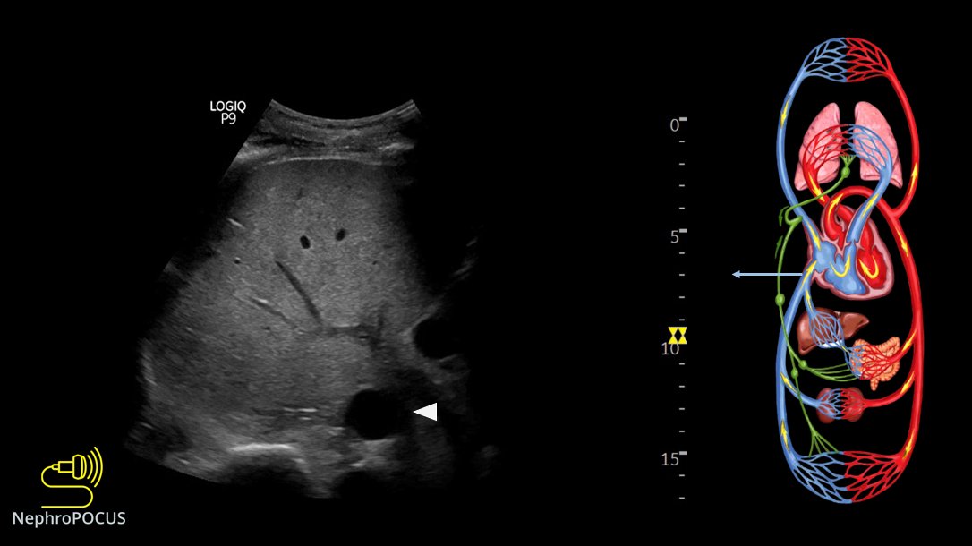
Here is the IVC👇
4/ 👆It is >2 cm, <50% collapsible. Definitely not consistent with a low right atrial pressure.
Here is hepatic vein #VExUS 👇
RAP meter shown on the top for beginners.
5/ 👆 Shows D-only pattern consistent with severe congestion.
There is not a lot of TR👇 Apical 4C shows bi-atrial enlargement (amyloidosis)
6/ Portal vein #VExUS👇
~40% pulsatility. Not terrible congestion yet, but getting there.
7/ Intra-renal #VExUS 👇
D-only pattern consistent with severe congestion. Color flow is great but not a happy kidney!
8/ Management: Strongly recommend to stop all the fluids (colloids/crystalloids). Hypotension is not due to hypovolemia. Consider drugs such as Droxidopa/pyridostigmine and maybe neurology consultation.
Take-home points:
1/How can #POCUS#echofirst help in the evaluation of #hyponatremia? #Nephrology consulted for low sodium. Pt received diuretic for shortness of breath (which was possibly due to hiatal hernia as shown on CXR).
2 physicians FELT patient was EUVOLEMIC
Small 🧵below 👇 #MedEd
2/Based on the above labs, it does look like euvolemic hyponatremia (Urine Na is high, Uosm >100 but not too high, BUN not high). BP was 150s systolic.
That's it? Give some salt tablets or UreNa etc.?
No; perform PHYSICAL EXAM (= #POCUS)
IVC was small - doesn't tell much 😬
3/ Next step? Look at the heart, assess stoke volume.
Recent echo LVEF>50%
LV contraction looks good visually
We measured LVOT VTI (couldn't measure LVOT diameter due to chest deformity precluding good PLAX view but its not a problem; VTI is generally enough) #POCUS
Time to discuss some rationale/evidence behind doing #VExUS#POCUS#Nephrology
A short #tweetorial#MedEd 👇 1/ Is fluid overload harmful?
of course yes. Here is a recent meta-analysis.
2/ Does fluid administration affect renal venous flow in asymptomatic but vulnerable patients (#heartfailure)? #POCUS#VExUS
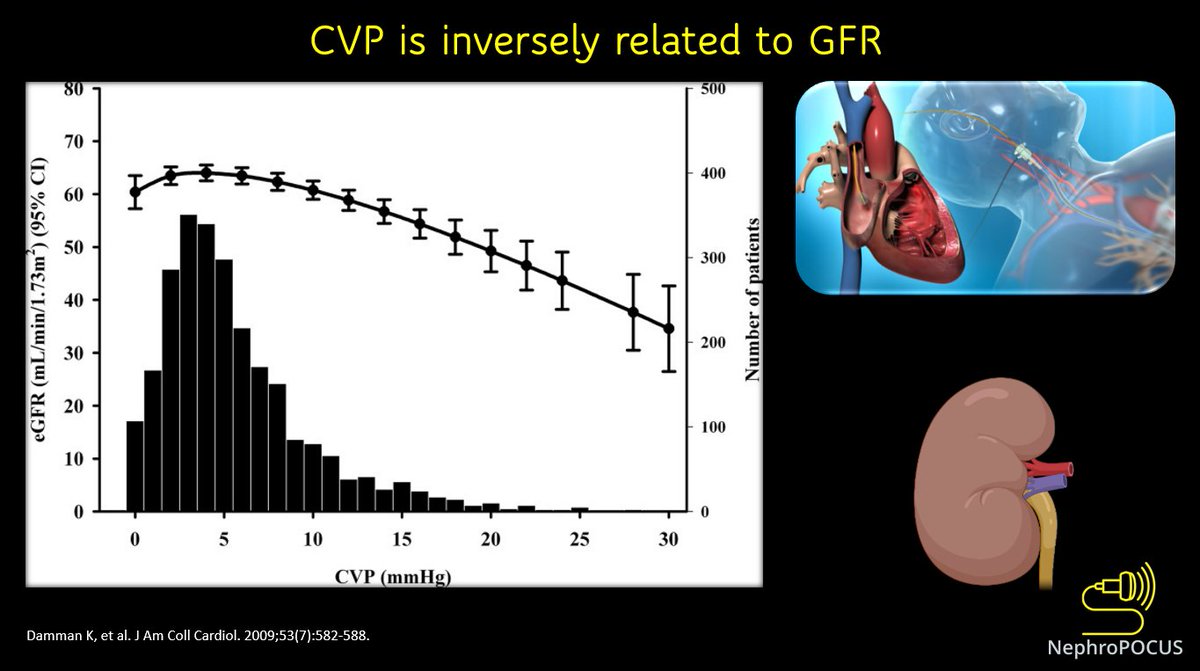
3/ In fact, elevated CVP is associated with reduced GFR.
This 👇is a study in outpatients undergoing right heart cath (N = 2557). In CVP values >6 mm Hg, a steep decrease in GFR was observed.
#POCUS#echofirst#MedEd case of the day.
Context: alcoholic liver cirrhosis with suspected #hepatorenal syndrome
Purpose: think about the underlying pathophysiology & seek expert input 1/ PLAX, PSAX, Apical views of the heart. Heart rate ~110-116 bpm #VExUS images in 🧵
2/ overall, looks hyperdynamic. LA looks little enlarged on PLAX, visual TAPSE high on apical.
In cirrhosis, hyperdynamic circulation is expected because of splanchnic vasodilatation and 'relative' arterial under filling.
LV EF #POCUS obtained using @kosmosplatform 👇
3/ Lets calculate stroke volume using LVOT VTI #POCUS
It looks supra-normal
OK #VExUS#POCUS enthusiasts, time for another case discussion.
Somebody asked if I ever recommend IV fluid in a patient with #VExUS 3.
Here is one example where I did. 1/ First, let's see the #physicalexam (#IMPOCUS) findings, then will tell about the case. #MedEd#Nephrology
2/ So, hepatic shows D-only pattern👆
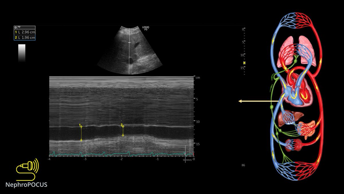
If we are doing #VExUS, IVC must be big. Here is the M-mode #POCUS 👇
3/ Renal parenchymal vein #VExUS#POCUS
It also demonstrates D-only pattern 👇
#POCUS quiz of the day:
Easy one. Identify 1, 2 and 3.
POLL in thread 👇 #IMPOCUS#MedEd
👆#POCUS
PE = pleural effusion
PER = pericardial effusion
Ao = aorta
IVC = inferior vena cava
Rt = right
Lt = left
Correct answer: option 2 - 1. left pleural effusion (note the appearance of collapsed lung; also u can see rib shadows/posterior chest wall = lung area) 2. Right pleural effusion (remember the Boomerang sign on subxiphoid view?) 3. IVC
Time for a #POCUS#tweetorial on optimization of Doppler. Very important for #VExUS enthusiasts. #MedEd 1/ Unlike greyscale imaging which depends on amplitude of the returned signal, Doppler depends on frequency information. This graphic explains why perpendicular angle is bad.
2/ other way of saying this, in relevance to color Doppler #POCUS
RBC moving away from the probe = Fr<Ft = negative Doppler shift = Blue color
RBC moving towards = Fr>Ft = positive Doppler shift = Red color
Rest of the images/videos from this excellent paper: pubs.rsna.org/doi/10.1148/rg… 3/ Anatomy (components) of a spectral Doppler waveform (carotid shown)👇
Above baseline is like red on color (towards probe), below = blue. As 0 degree angle is not always possible, <60 is considered OK.