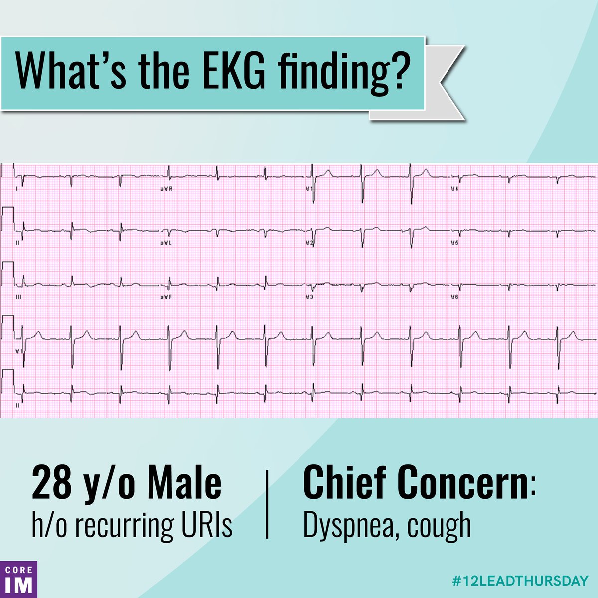
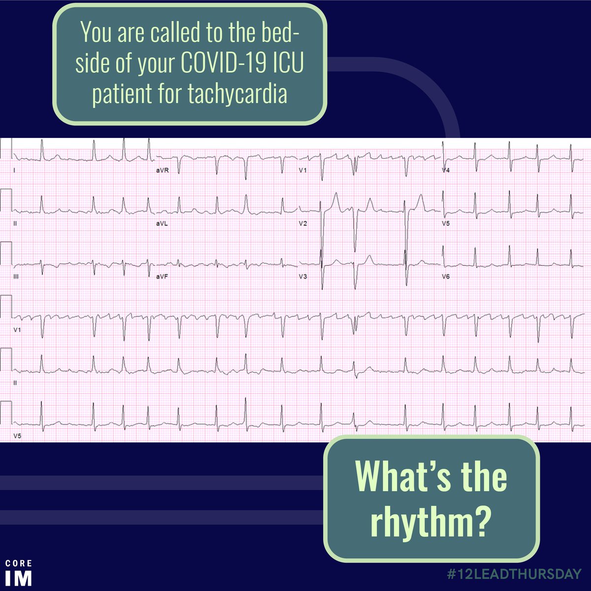
1/ Good morning #MedTwitter, it’s time for another episode of #12LeadThursday! Remember to approach every EKG systematically. Grab your calipers, and let’s dive in!

What are potential causes of this pause?
2/ We can think about pauses in three buckets below. We’ll get into why we think a PAC is causing the pause above, but stop for a moment and consider: what would the EKG look like if AVN blockade or sinus node dysfunction were at play?
3/ In the above EKG, we see the PAC hiding in a T wave! This PAC reset the SA node, and a pause was born!

Before we move on: if the AVN is dysfunctional, how do you differentiate a blocked PAC from a dropped beat?
4/ In AV block, the P-P interval is constant. The single or intermittently dropped beats occur at regular conduction intervals such as 3:2, 4:3, 5:4.

For non-conducted PACs, the P-P is altered, but not consistent.
5/ Huge thanks to the author of this byte, @ArisSikolas, and as always to the co-directors of #12LeadThursday: @KelseyGrossman and @GregoryKatz.

You can find this byte and more on our website: bit.ly/3Bf4NE1

• • •

Missing some Tweet in this thread? You can try to force a refresh
 

Keep Current with CORE IM

CORE IM Profile picture

Stay in touch and get notified when new unrolls are available from this author!

Read all threads

This Thread may be Removed Anytime!

PDF

Twitter may remove this content at anytime! Save it as PDF for later use!

Try unrolling a thread yourself!

how to unroll video
  1. Follow @ThreadReaderApp to mention us!

  2. From a Twitter thread mention us with a keyword "unroll"
@threadreaderapp unroll

Practice here first or read more on our help page!

More from @COREIMpodcast

24 Jun
1/ Good morning, #medtwitter, and happy Thursday! We’re excited to bring you our latest edition of #ReadingRoom. Dim the lights, and get ready to clinically correlate!

You’ve got two abdominal plain films below. Which one is from a patient with a SBO?
2/ Let’s take a closer look, starting with the image on the right: here, there’s no abnormal dilation and the distribution of gas is normal. This is a normal abdominal film.
3/ On the other hand, here we see an abnormally dilated small bowel with predominant small bowel gas while there is no rectal gas and scant colonic gas. This is concerning for a SBO.
Read 7 tweets
13 Apr
1/ Good morning, #MedTwitter, and happy Tuesday! We’re excited to bring you our latest installment of #ReadingRoom today. Dim the lights; it’s time to clinically correlate!

What object is labeled in the CXR below? What is it used for? Image
2/ That, friends, is our dear friend: the NG tube. It is our portal for tube feeds and PO meds (technically Per Nasus?), or if you put the thing down flip it and reverse it you can use the NGT to decompress the stomach. Image
3/ Next question: should you place that order that says “OK to use NG tube”?

The graphic below shows a systematic approach to ensure correct placement. This one is good to go! Image
Read 6 tweets
25 Mar
1/ #MedTwitter Today, March 25, we want to remember the Medical Committee for Human Rights (MCHR), a branch of the Civil Rights movement. Image
2/ MCHR recognized the fundamental inseparability of fight for racial justice and the fight for health equity. Image
3/ MCHR no longer exists in name, but it lives on in the legacy of institutions like @PNHP and @P4HR.

So, #MedTwitter, consider this a call to action: what are you doing to promote equity in healthcare today? Image
Read 4 tweets
13 Aug 20
1/ It’s Thursday, #MedTwitter, which means it’s time to take out your calipers and get to business. It’s #12LeadThursday!

Remember to use your system to go through each and every ECG methodically.
2/ Rate: ~75bpm
Rhythm: NSR
Axis: ...
3/ Do you have a schema or framework for thinking about a right axis deviation? What’s your differential?

Let us know in the comments below, and check out the framework we’ve provided!
Read 7 tweets
16 Jul 20
1/ Welcome back, #medtwitter, to the latest installment of #12LeadThursday - #covid19 edition! Let’s put those calipers to work!

Take a look at this ECG. Approach it systematically. What do you see?
2/ In the above ECG, you’ve got an irregular narrow complex tachycardia. What’s your final read? Reply with your reasoning!
3/ Let’s take a closer look. In the graphic below, we’ve highlighted what you would expect to see in flutter - and none of it is present here. Our patient is fibbin’ away, folks.
Read 6 tweets
14 Jul 20
1/ Happy Tuesday, #medtwitter! It’s time to take that walk down the hall to radiology for another round of #readingroom - let’s go!
2/ Looking at the previous tweet, where is the abnormal air located?
3/ If you said mediastinum, you’re right! This is also well characterized on chest CT pictured below.

Next question: how on earth did air get into the mediastinum? Reply with your thoughts, differentials and/or schema!
Read 7 tweets

Did Thread Reader help you today?

Support us! We are indie developers!


This site is made by just two indie developers on a laptop doing marketing, support and development! Read more about the story.

Become a Premium Member ($3/month or $30/year) and get exclusive features!

Become Premium

Too expensive? Make a small donation by buying us coffee ($5) or help with server cost ($10)

Donate via Paypal Become our Patreon

Thank you for your support!

Follow Us on Twitter!

:(