A review of upper extremity Eponym Fractures.
It is important for clinicians to be aware of eponymous fractures as they are commonly used and allow for a succinct description of sometimes complex injuries.
🦴⚒️🧵👇
To view our past reviews, visit:
💻 myorthoreviews.com📱
It is important for clinicians to be aware of eponymous fractures as they are commonly used and allow for a succinct description of sometimes complex injuries.
🦴⚒️🧵👇
To view our past reviews, visit:
💻 myorthoreviews.com📱

First off, what is an eponym fracture?
Eponymous: of, relating to, or being the person or thing for whom or which something is named.
Eponym fractures are named based on the first person to describe them (e.g. Holstein-Lewis) or by an activity (e.g. Chauffer's)
Eponymous: of, relating to, or being the person or thing for whom or which something is named.
Eponym fractures are named based on the first person to describe them (e.g. Holstein-Lewis) or by an activity (e.g. Chauffer's)
We will review the eponymous fractures of the upper extremity going from proximal to distal.
To start off, what is an eponym for the following lesion?
To start off, what is an eponym for the following lesion?


This is an example of a Hill-Sachs lesion.
These occur during anterior shoulder dislocation as the posterolateral humeral head impacts the anterior glenoid rim.
Larger lesions may engage the glenoid and require repair.
On the contrary, what is an eponym for this lesion?
These occur during anterior shoulder dislocation as the posterolateral humeral head impacts the anterior glenoid rim.
Larger lesions may engage the glenoid and require repair.
On the contrary, what is an eponym for this lesion?


This is an example of a Reverse Hill-Sachs lesion.
These occur during posterior shoulder dislocation as the anteromedial humeral head impacts against the posterior glenoid rim.
What is an eponym for this fracture?
These occur during posterior shoulder dislocation as the anteromedial humeral head impacts against the posterior glenoid rim.
What is an eponym for this fracture?

This is an example of a Bony-Bankhart lesion
During anterior shoulder dislocation an injury to the anteroinferior labrum may occur, a Bankhart Lesion. An avulsion fracture of the anteroinferior glenoid may also occur, a Bony-Bankhart Lesion
These can lead to chronic instability
During anterior shoulder dislocation an injury to the anteroinferior labrum may occur, a Bankhart Lesion. An avulsion fracture of the anteroinferior glenoid may also occur, a Bony-Bankhart Lesion
These can lead to chronic instability
What is an eponym for the following fracture?

This is an example of a Holstein-Lewis Fracture.
A spiral fracture of the distal ⅓ humeral shaft. This fracture pattern is associated with an increased risk of radial nerve injury.
A spiral fracture of the distal ⅓ humeral shaft. This fracture pattern is associated with an increased risk of radial nerve injury.
This patient has a comminuted fracture of the radial head and dislocation of the distal radioulnar joint.
What is an eponym for the following fracture?
What is an eponym for the following fracture?

This is an example of an Essex-Lopresti fracture-dislocation.
These injuries are rare and commonly missed initially (as high as 80%) and an isolated radial head fracture should prompt evaluation of the DRUJ.
What is an eponym for the following fracture?
These injuries are rare and commonly missed initially (as high as 80%) and an isolated radial head fracture should prompt evaluation of the DRUJ.
What is an eponym for the following fracture?

This is an example of a Monteggia fracture-dislocation.
This is a fracture of the proximal ulnar shaft with dislocation of the radiocapitellar joint.
What is an eponym for the following fracture?
This is a fracture of the proximal ulnar shaft with dislocation of the radiocapitellar joint.
What is an eponym for the following fracture?

This is an example of a Galeazzi fracture-dislocation.
It is a fracture of the distal radial shaft with disruption of the DRUJ. Of note, DRUJ disruption may be subtle.
Radial fractures within 7.5 cm of the wrist are associated with higher rates of chronic instability.
It is a fracture of the distal radial shaft with disruption of the DRUJ. Of note, DRUJ disruption may be subtle.
Radial fractures within 7.5 cm of the wrist are associated with higher rates of chronic instability.

What is an eponym for this fracture?

This is an example of a night-stick fracture.
It is an isolated ulnar shaft fracture secondary to direct trauma.
It is an isolated ulnar shaft fracture secondary to direct trauma.

Now onto two of the most well-known eponym fractures, that are also very easily confused.
What are the eponymous names of the following fractures?
What are the eponymous names of the following fractures?
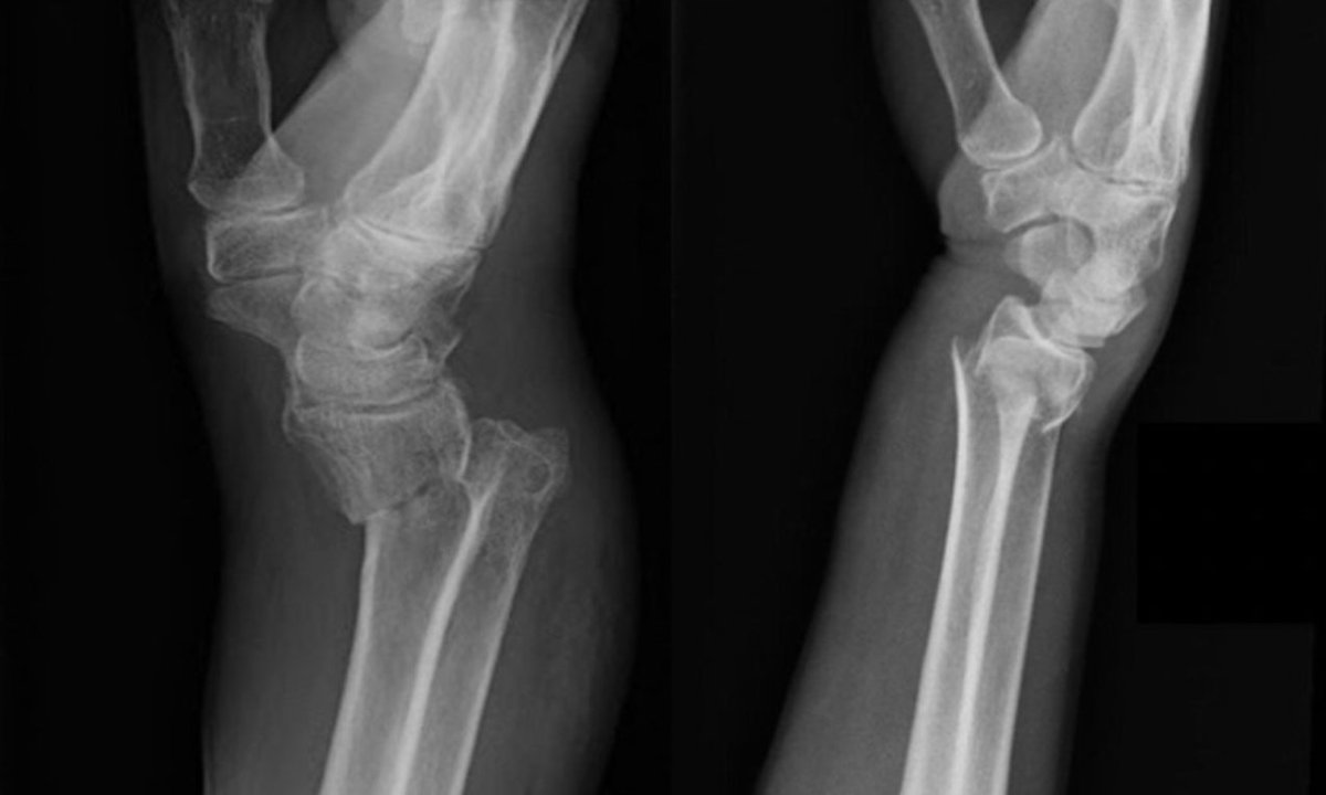
On the left, we have a Smith's Fracture.
These are extraarticular distal radius fractures with volar displacement.
On the right, we have a Colles' Fracture.
These are extraarticular distal radius fractures with dorsal displacement.
These are extraarticular distal radius fractures with volar displacement.
On the right, we have a Colles' Fracture.
These are extraarticular distal radius fractures with dorsal displacement.

What are the eponymous names for the following fractures?
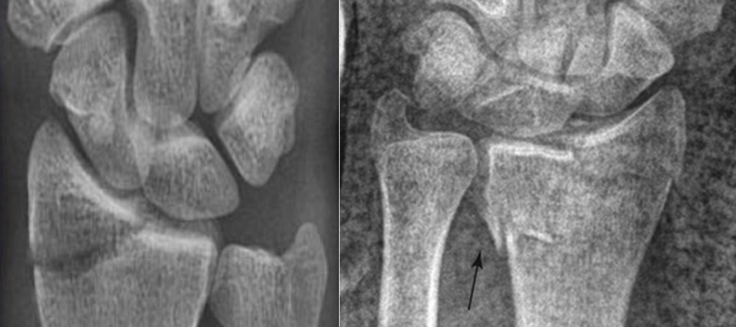
On the left, we have a Chauffer's fracture. This is a radial styloid fracture caused by impaction against the scaphoid.
On the right, we had a Die-punch fracture. This is an intra-articular fracture of the lunate fossa caused by axial loading through the lunate.
On the right, we had a Die-punch fracture. This is an intra-articular fracture of the lunate fossa caused by axial loading through the lunate.

What is an eponym for the following fracture?

This is an example of a Volar Barton's Fracture.
These can involve either the volar or dorsal lip of the distal radius.
Some may refer to a Dorsal Bartons as a "Barton's fracture" and Volar Bartons as a "Reverse Barton's fracture.
What is an eponym of the following fracture?
These can involve either the volar or dorsal lip of the distal radius.
Some may refer to a Dorsal Bartons as a "Barton's fracture" and Volar Bartons as a "Reverse Barton's fracture.
What is an eponym of the following fracture?

This is an example of a Boxer's Fracture.
These occur when an untrained person strikes with a flexed wrist.
Professional fighters would most likely fracture their 2nd or 3rd metacarpal because they would strike with a neutral wrist.
These occur when an untrained person strikes with a flexed wrist.
Professional fighters would most likely fracture their 2nd or 3rd metacarpal because they would strike with a neutral wrist.

What are the eponymous names of the following metacarpal base fractures?

On the left is a Bennett's Fracture. This fracture may be displaced by the abductor pollicis longus.
In the middle is a Rolando Fracture, a comminuted fracture of the 1st metacarpal base.
On the right is a Reverse or Baby Bennet Fracture, this may be displaced by the ECU.
In the middle is a Rolando Fracture, a comminuted fracture of the 1st metacarpal base.
On the right is a Reverse or Baby Bennet Fracture, this may be displaced by the ECU.

If you enjoyed this review please like or retweet to help the page grow and give us a follow.
Author: @CSMorford
#EponymFractures #UpperExtremity #Trauma #OrthoTwitter #MedED #DistalRadius #Fractures #MedTwitter #Trauma #Orthopedics #Tweetorials
Author: @CSMorford
#EponymFractures #UpperExtremity #Trauma #OrthoTwitter #MedED #DistalRadius #Fractures #MedTwitter #Trauma #Orthopedics #Tweetorials
• • •
Missing some Tweet in this thread? You can try to
force a refresh