2/A “syndromic appearing” young adult pt who was a poor historian & could not specify any prior diagnosis, p/w left neck swelling. On CTA, calling the IJ supersized would have been an understatement
3/Posterior to the IJ was a tangle of vessels, but no identifiable soft tissue mass, concerning for a vascular malformation. Catheter angiography showed a Jackson Pollack painting appearance of tangled vessels consistent with an AVM
4/But it was more complicated than that. Although there was an AVM, there were also signs of a low flow lesion as well. There was non-enhancing soft tissue & phleboliths that looked more like a venolymphatic. But an enlarged main pulmonary trunk indicated a high flow lesion.
5/And among the vascular malformation was all this extra fat. It didn’t look like an encapsulated lipoma. It was more like just overgrowth of the fat in this region—don’t we all have problems with a little bit of fatty overgrowth! 😉
6/An MRI of the brain showed a Chiari 1 and bright spots in the cerebellum that looked like the UBO (unidentified bright objects) one sees in neurofibromatosis 1 pts. But this patient had no other stigmata of NF1.
7/So we have a vascular malformation (mixed high & low flow) & lipomatous overgrowth. This is CLOVES syndrome (Congenital Lipomatous Overgrowth w/combined-type Vascular malformations, Epidermal naevi, Skeletal anomalies). They can also have posterior fossa abnormalities.
8/CLOVES actually falls under the umbrella of a spectrum of vascular abnormalities/lipomatous overgrowth syndromes—the most famous being Proteus syndrome—the syndrome the elephant man had. I never thought I would come across a disease that is a variant of the elephant man!
9/So next time you see a vascular malformation & lipomatous overgrowth—think of this umbrella of PROS syndromes—even if you are an adult neuroradiologist like me who NEVER sees such syndromes (real life picture of me below every time pediatric pathology comes across my screen)
• • •
Missing some Tweet in this thread? You can try to
force a refresh
2/The hardest part of a pituitary MRI is deciding if there is cavernous sinus invasion. It makes sense that the more lateral a tumor goes on MRI, the more likely it has invaded the sinus—bc it is going the direction of the sinus. But how far is far enough?
3/This is important bc each time a radiologist makes a call on imaging, they make a bet & they are betting their credibility. And unlike other bets, there is only 1 wager—all in! So it is important to not call it when you might be wrong, bc overcalls destroy credibility.
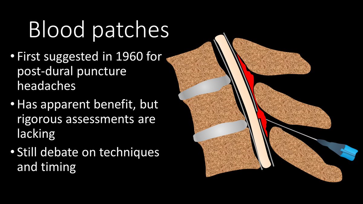
1/”Now your mouth will drop when you see the cord compression we caused,” I said to my fellow looking at our targeted #bloodpatch CT, “But take a deep breath—that’s actually what we want.”
A #tweetorial about CSF leaks & blood patches! #medtwitter#CSFleak#neurotwitter#neurorad
2/Epidural blood patches (EBPs) have been around since the 60s. Blood was first injected in the epidural space to try to plug the leak in post-dural puncture HA. It has now been expanded to other CSF leaks. However, controlled studies are lacking & therefore methods vary greatly
3/No one is sure of how EBPs work. Some believe blood directly plugs the leak site. Other believe it’s a pressure effect--injected blood increases epidural pressure, squeezing the thecal sac like a stress ball, elevating subarachnoid CSF pressure to relieve low pressure HA.
2/Sphenopalatine ganglion (SPG) is the largest collection of neurons outside the brain—like a mini brain just for your face. It contains sensory, sympathetic, & parasympathetic nerve fibers. Given this, it’s not surprising that it’s felt to contribute to facial pain syndromes
3/SPG is a meeting point for the sensory nerves from V2 (thus related to trigeminal neuralgia) & the sympathetics and parasympathetics from the greater superficial and deep petrosal nerves, which have been implicated in cluster headache, migraine, & other facial pain syndromes.
2/CT in acute stroke has 2 main purposes—(1) exclude intracranial hemorrhage (a contraindication to thrombolysis) & (2) exclude other pathologies mimicking acute stroke. However, that doesn’t mean you can’t see other findings that can help you diagnose a stroke.
3/Infarct appearance depends on timing. In first 12 hrs, the most common imaging finding is…a normal head CT. However, in some, you see a hyperdense artery or basal ganglia obscuration. Later in the acute period, you see the insular ribbon & sulcal effacement
2/Its name “hippocampus” comes from its shape on gross anatomy. Early anatomists thought it looked like an upside down seahorse—w/its curved tail resembling the tail of a seahorse. Hippocampus literally means seahorse.
3/In cross section, it has a spiral appearance, leading to its other name, Cornu Ammonis, translated Ammon’s Horn. Ammon was an Egyptian god w/spiraling rams horns. The hippocampal subfields are abbreviated CA-1, CA-2, etc, w/CA standing for “Cornu Ammonis”
2/We’ll talk about the imaging part of TLICS. TLICS scores a fx on (1) morphology & (2) posterior ligamentous complex (PLC) injury. Let’s start w/morphology. W/only mild axial loading, you get the simplest fx, a compression fx—like a simple long bone fx--worth 1 pt.
3/As the axial force grows, this becomes a burst fx with retropulsion of the posterior vertebral body—just as greater force causes more comminution in long bone fxs. A burst is worth 2 points.