2/Here are 3 lesions, all round and bright and in the region of the foramen of Monro. Can you tell from the images which is a colloid cyst and which may be something else? Choose which one or ones you think are a colloid cyst
Choose which one you think is a colloid cyst
4/In this case it was A. B was a tortuous basilar and C was a cavernoma of the chiasm/hypothalamus that had bled and projected into the third ventricle.
5/Many lesions may mimic a colloid cyst at the foramen of Monro. Below is a list, but it is by no means exhaustive. So with so many mimickers, how can you know when to call a colloid cyst?
6/They say location is everything--especially in colloid cysts. 99% of them are located at the foramen of Monro, so if it isn't at the foramen, be suspicious that it isn't a colloid cyst
7/Another feature that makes it special is actually how few special features it has! It should be very featureless. Many imaging findings we use to characterize lesions (enhancement, calcification, diffusion restriction), should all be absent in a colloid cyst
8/I remember this bc colloid cysts are kind of cousins to other midline congenital cysts (Rathke's cyst & Thornwaldt cyst) & they behave similarly. So if there's a feature that would be weird in a Rathke's or Thornwald cyst (calcs, enhancement), it's weird for a colloid cyst
9/But recognizing a colloid cyst isn't enough. There are important things to mention in your report. You should mention anatomic variants of the septum & fornix that could affect the surgical approach. Also mention low T2 signal, as these cysts can be more difficult to resect
10/Another important issue is where along the 3rd ventricle the cyst extends. Zone 1 is anterior to the mass intermedia, Zone 2 is behind Zone 1 but anterior to the aqueduct, and Zone 3 is behind Zone 2. Zones 1 & 3 are higher risk
11/I hate it when classifications don't go in order. I want Zone 1 to be lowest risk and Zone 3 highest. I hate it when there is a sine wave of risk in the classification
12/But you can remember this by remembering that there are openings at the anterior & posterior 3rd ventricle. So anteriorly you are at risk of obstructing the foramen & posteriorly the aqueduct. Zone 2 is just the zone sandwiched between to the two openings, so it is low risk.
13/So remember, there are mimics of colloid cysts all around. So look at the imaging findings, instead of listening to the siren song!
• • •
Missing some Tweet in this thread? You can try to
force a refresh
2/Some believe that the peaked, mountain like appearance of the frontal lobes is a critical sign of a life-threatening complication & should be called & reported. Others believe it is too non-specific, is commonly seen when there isn’t tension & should be retired. Who’s right?
3/First, let’s clarify about what the Mt Fuji sign actually is. Most are familiar with the fact that large collections of pneumocephalus can compress the frontal lobes—making them look like the slopes of a mountain. But this isn’t actually enough to call Mt Fuji.
2/A “syndromic appearing” young adult pt who was a poor historian & could not specify any prior diagnosis, p/w left neck swelling. On CTA, calling the IJ supersized would have been an understatement
3/Posterior to the IJ was a tangle of vessels, but no identifiable soft tissue mass, concerning for a vascular malformation. Catheter angiography showed a Jackson Pollack painting appearance of tangled vessels consistent with an AVM
2/The hardest part of a pituitary MRI is deciding if there is cavernous sinus invasion. It makes sense that the more lateral a tumor goes on MRI, the more likely it has invaded the sinus—bc it is going the direction of the sinus. But how far is far enough?
3/This is important bc each time a radiologist makes a call on imaging, they make a bet & they are betting their credibility. And unlike other bets, there is only 1 wager—all in! So it is important to not call it when you might be wrong, bc overcalls destroy credibility.
1/”Now your mouth will drop when you see the cord compression we caused,” I said to my fellow looking at our targeted #bloodpatch CT, “But take a deep breath—that’s actually what we want.”
A #tweetorial about CSF leaks & blood patches! #medtwitter#CSFleak#neurotwitter#neurorad
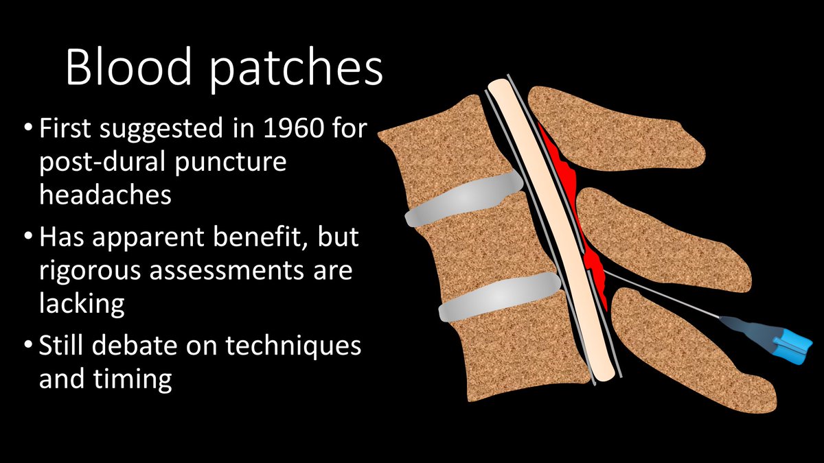
2/Epidural blood patches (EBPs) have been around since the 60s. Blood was first injected in the epidural space to try to plug the leak in post-dural puncture HA. It has now been expanded to other CSF leaks. However, controlled studies are lacking & therefore methods vary greatly
3/No one is sure of how EBPs work. Some believe blood directly plugs the leak site. Other believe it’s a pressure effect--injected blood increases epidural pressure, squeezing the thecal sac like a stress ball, elevating subarachnoid CSF pressure to relieve low pressure HA.
1/”You want me to put my needle where??” my fellow asked incredulously. It’s daunting, but it works—image-guided sphenopalatine ganglion blocks
Here’s a #tweetorial about this underutilized but effective procedure! #medtwitter#neurology#headache#migraine#neurotwitter#FOAMed
2/Sphenopalatine ganglion (SPG) is the largest collection of neurons outside the brain—like a mini brain just for your face. It contains sensory, sympathetic, & parasympathetic nerve fibers. Given this, it’s not surprising that it’s felt to contribute to facial pain syndromes
3/SPG is a meeting point for the sensory nerves from V2 (thus related to trigeminal neuralgia) & the sympathetics and parasympathetics from the greater superficial and deep petrosal nerves, which have been implicated in cluster headache, migraine, & other facial pain syndromes.
2/CT in acute stroke has 2 main purposes—(1) exclude intracranial hemorrhage (a contraindication to thrombolysis) & (2) exclude other pathologies mimicking acute stroke. However, that doesn’t mean you can’t see other findings that can help you diagnose a stroke.
3/Infarct appearance depends on timing. In first 12 hrs, the most common imaging finding is…a normal head CT. However, in some, you see a hyperdense artery or basal ganglia obscuration. Later in the acute period, you see the insular ribbon & sulcal effacement