HYPOTHALAMUS (HT)🧵-the control center of circadian rhythm, fatigue/wakefulness, hunger/satiety, sex drive, thirst/BP—and the command post for endocrine control via the HT-pituitary axis.
#meded #neuroradiology #neuroscience #radiology #neurology #neurosurgery #neuroanatomy
1/28
#meded #neuroradiology #neuroscience #radiology #neurology #neurosurgery #neuroanatomy
1/28
Quiz A: Histamine activity in the brain/brainstem contributes to alertness/wakefulness–hence: sleepy effects of benadryl. Histaminergic neurosecretory cell bodies are exclusively in the tuberomammillary nucleus of the TUBER CINEREUM (HT floor), with widespread projections.
2/28
2/28

The HT is a mysterious and complex—I get confused sighs from medical students when the topic arises—the small almond-sized morsel is the control center for endocrine/hormone regulation, and homeostasis of food/water consumption, sleep, BP, sex/attachment. The HT is boss!
3/28
3/28
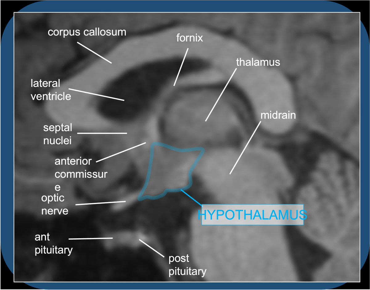
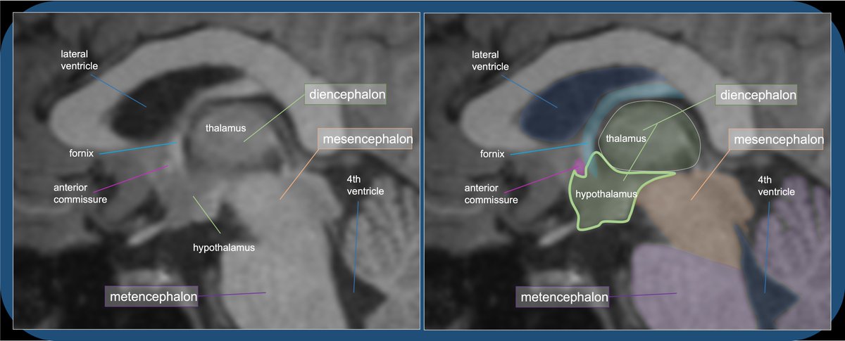
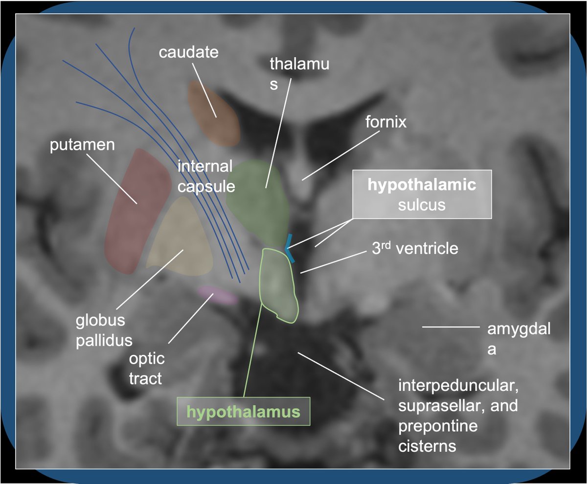
Where is it? The HT is at the ventral diencephalon, a diamond-shaped collection of nuclei and fiber tracts just anterior/inferior to the thalamus.
4/28
4/28
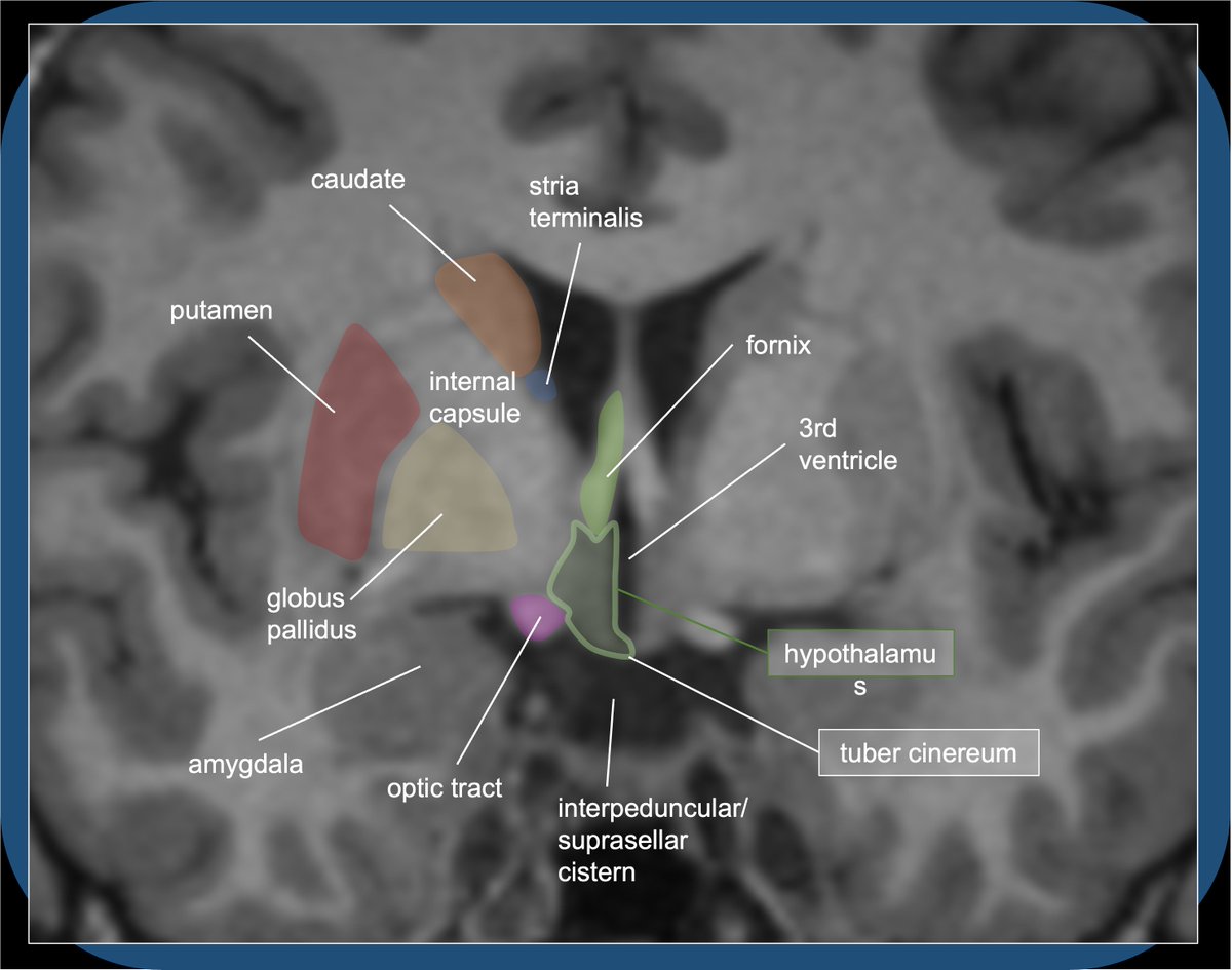
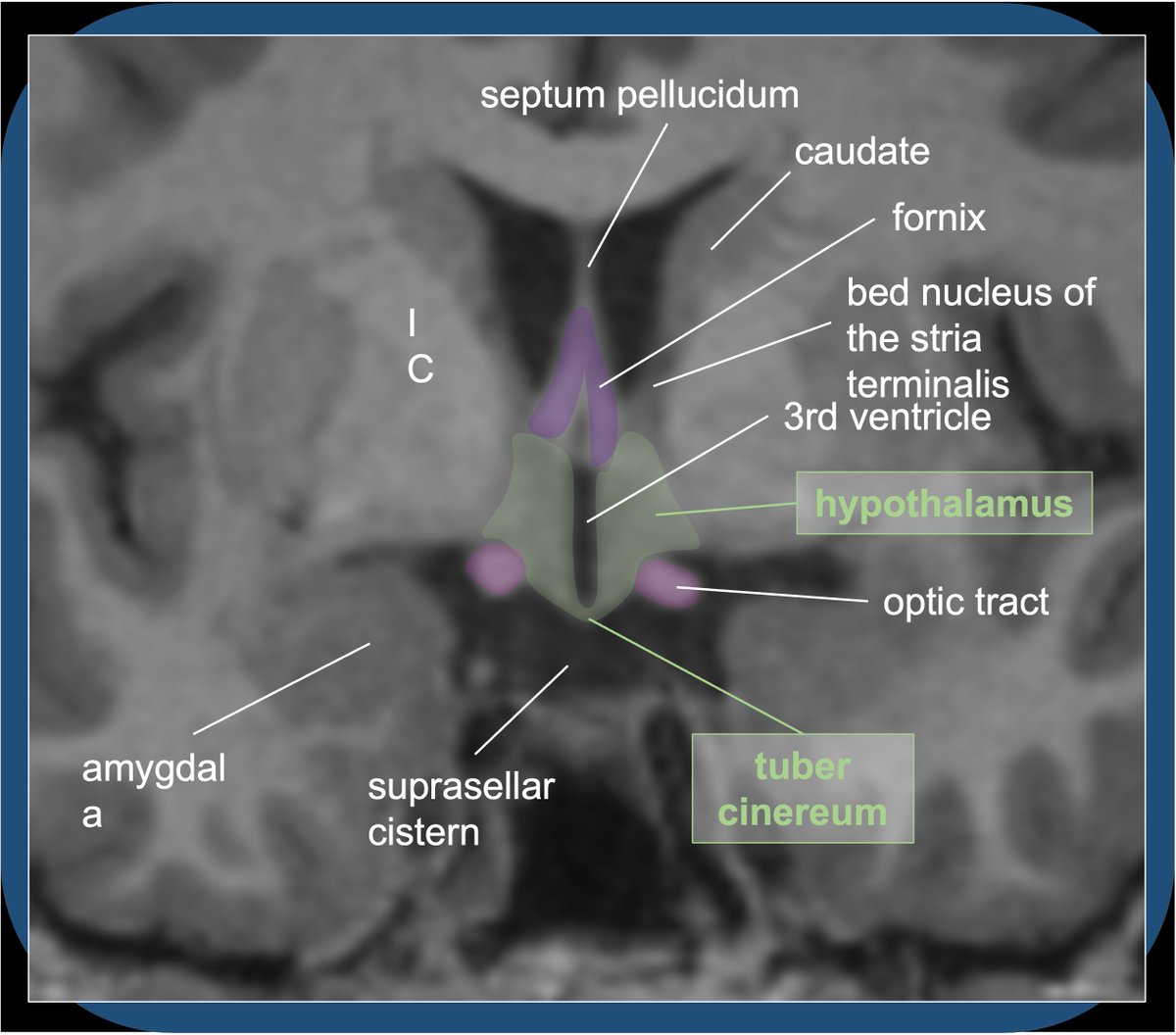
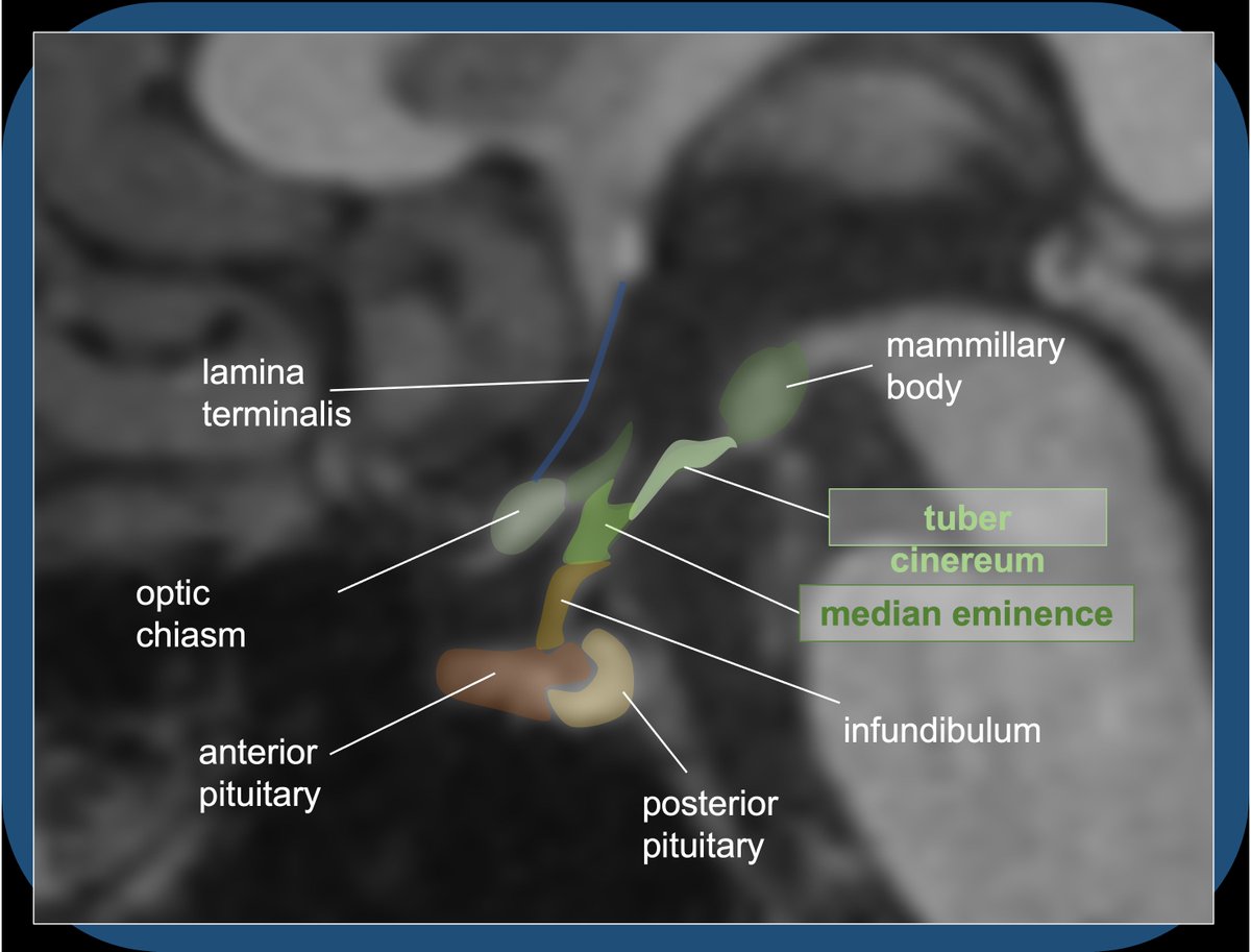
There are 2 halves to the HT, on either side of a recess of the anterior 3rd ventricle. The 2 halves meet in the midline inferiorly, forming the floor of the hypothalamus--the TUBER CINEREUM (TC).
5/28
5/28
The HYPOTHALAMUS roughly extends to the ANTERIOR COMMISSURE at the anterior/superior margin, LAMINA TERMINALIS anterior, and OPTIC CHIASM/TRACT at the anterior/inferior margin.
6/28
6/28

The sup margin borders the thalamus (separated by the HYPOTHALAMIC SULCUS, a groove in lateral wall of 3rd ventricle). Post margins are the mammillary bodies and midbrain peduncles. Inferior margin is the basal cisterns. Lateral margin borders int capsule and optic tracts.
7/28
7/28
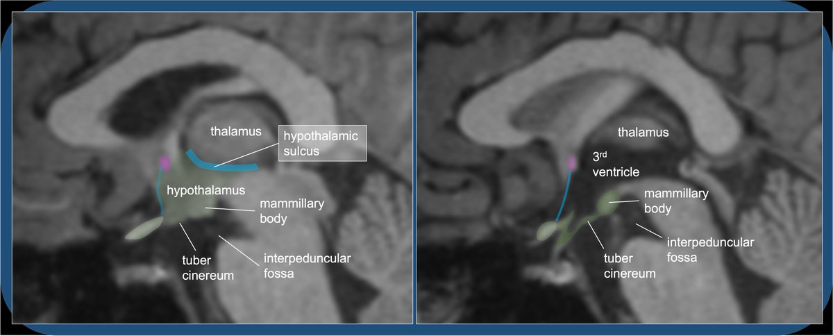
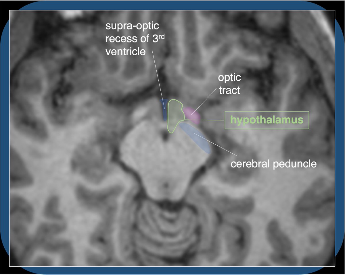
Medially, the 3rd ventricle separates the two halves of the HT; SUPRAOPTIC and INFUNDIBULAR RECESSES extend ant/inferior. Note—it can be difficult to see lamina terminalis separating 3rd ventr. recesses from subarachnoid space of basal cisterns, which border HT anteriorly.
8/28
8/28


The TUBER CINEREUM (TC) is a median protuberance between optic chiasm (ant) and mammillary bodies (post), forming the floor of the hypothalamus, and bordering the INTERPEDUNCULAR & SUPRASELLAR basal cisterns (as well as the cistern of the lamina terminalis).
9/28
9/28
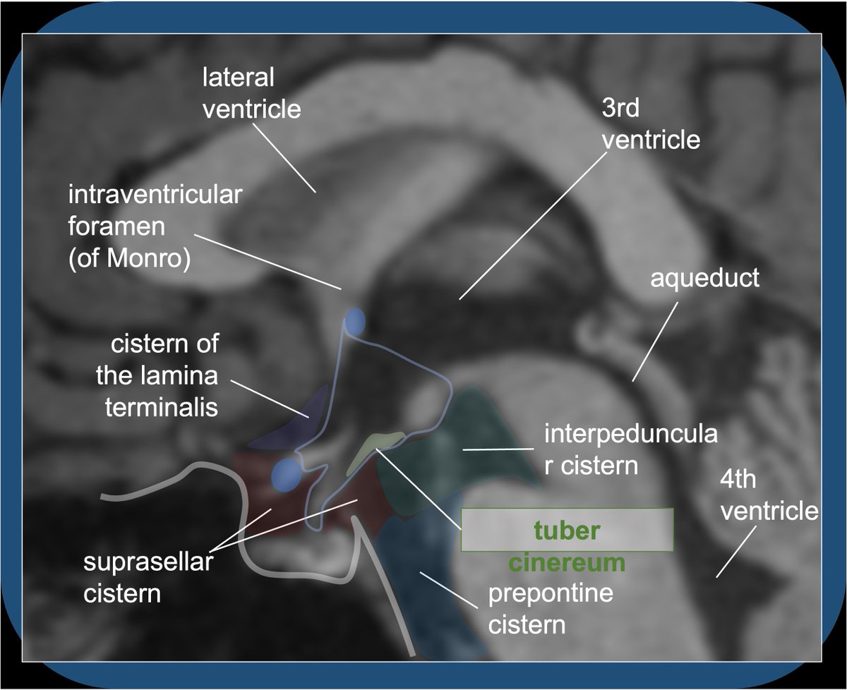
At the anterior margin of the tuber cinereum, the MEDIAN EMINENCE is a small swelling at the base of the pituitary stalk/infundibulum—it is special because it lacks a BBB, allowing for release of HT hormones into the hypophyseal portal system.
10/28
10/28
The many HT nuclei are not distinguished on imaging, and can be subdivided in various ways, e.g. anterior/chiasmatic (ACA/Acomm); median/tuberal (PCA); post/mammillary (Pcomm/PCA/BA). Nuclei are grouped variably depending on source (e.g. is PV middle or ant?) Semantics…
11/28
11/28

Subdivisions can be further divided into 3 morphologic/functional areas (lateral, medial, periventricular). The fornix and mammillothalamic tracts split the HT into medial and lateral parts.
12/28
12/28

Speaking of...the white matter of the FORNIX and MAMMILLOTHALAMIC TRACT are well seen on T1 images, descending/ascending amid the gray nuclei of the HT.
13/28
13/28

14/28
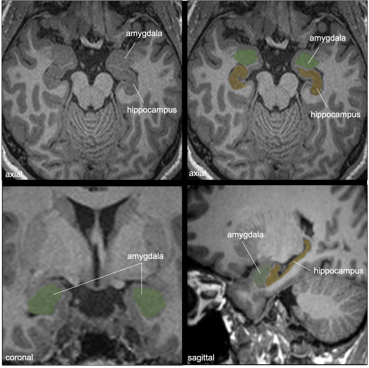
These tracts are part of the Papez circuit (discussed in a previous hippocampal tweetorial:
15/28
https://twitter.com/aaronrutman/status/1315906265294401542)--important in episodic memory formation—inputs from hippocampus➡️MBs via fornix; subsequent input➡️anterior nucleus of thalamus via mammillothalamic tract.
15/28

The mammillary bodies, usually categorized with the HT, but functionally a limbic structure, are a pair of dome-shaped structures at the undersurface of the diencephalon (at the posterior margin of the tuber cinereum). Damage results in episodic memory difficulties.
16/28
16/28
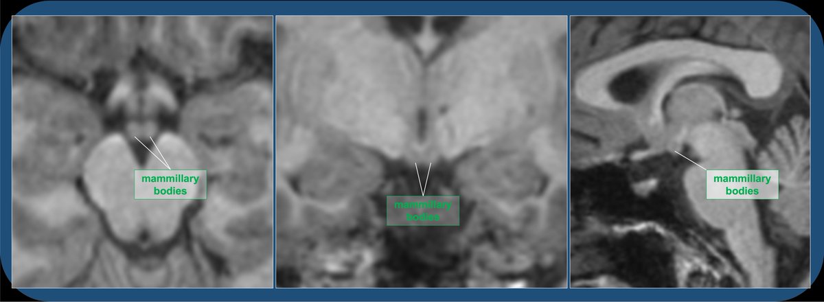
In Wernicke-Korsakoff Encephalopathy, vitamin B1 deficiency (often assoc with alcoholism) damages the mammillary bodies, parts of the thalamus, and periaqueductal gray (among others), leading to both anterograde and retrograde memory impairment. cases.rsna.org/fullscreen-ima…
17/28
17/28

The many other HT nuclei have overlapping functions and most are not possible to identify with imaging except by estimating location; but a quick rundown of functions is useful and interesting.
18/28
18/28
The suprachiasmatic nucleus recieves photic afferents from the retina via the retinohypothalamic tract, and acts as the dominant regulator of circadian rhythm.
19/28
19/28

Hunger and satiety are mainly controlled by the dorsomedial and ventromedial nuclei respectively.
20/28
20/28
Thermoregulation is mainly controlled by the posterior nucleus (heating) and anterior nucleus (cooling).
21/28
21/28
Systemic osmotic balance is controlled by the supraoptic and paraventricular nuclei (vasopressin, oxytocin); paraventricular also important for pituitary stimulating hormone production (CRH, TRH, GnRH, GHTH).
22/28
22/28
Medial preoptic (a sexually dimorphic nucleus, larger in males), regulates sex hormones and behavior.
23/28
23/28
Lateral preoptic is involved in sleep onset (via orexin), and also helps regulate hunger. Clinical note: narcolepsy is a neurodegenerative disorder affecting orexin producing cells of the lateral hypothalamus (which can be catalyzed by infectious & autoimmune processes).
24/28
24/28
The arcuate nucleus, which is horseshoe-shaped and overlies the median eminence at the lateral/caudal root of the infundibulum, produces GHRH & dopamine to affect the H-P axis, and provides projections to help regulate hunger, sex drive, etc.
25/28
25/28

And so much more detail to know—more nuclei, hormones, efferent & afferent tracts, etc. The cellular and functional anatomic details may be less important for the average radiologist, surgeon, or oncologist (probably good to know for neurologist or endocrinologist!)…
26/28
26/28
…but a neurorad should know the anatomy and how to describe the hypothalamic & limbic structures that may be affected by sellar tumors (craniopharyngiomas, hamartomas, pit adenomas, germinoma, mets, optic gliomas), or toxic, metabolic, or inflammatory encephalopathies.
27/28
27/28

Hypothalamic hamartomas (a benign non-neoplastic heterotopia), may be sessile involving MBs, or pedunculated/exophytic from tuber cinereum. They are associated with gelastic seizures in kids.
Case courtesy of Aruna Pallewatte, Radiopaedia.org, rID: 28460
28/28
Case courtesy of Aruna Pallewatte, Radiopaedia.org, rID: 28460
28/28

• • •
Missing some Tweet in this thread? You can try to
force a refresh