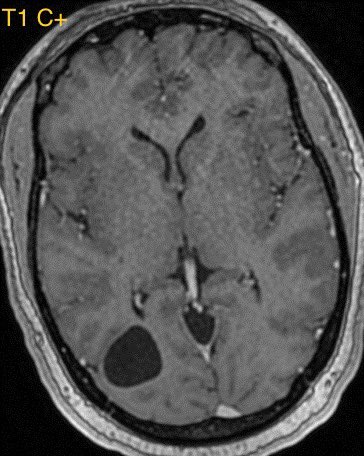
What is the most likely diagnosis in this 25 y/o M with headache? 🧠

Answer later tonight #radres #Neurology #Neurosurgery #MedEd #MedTwitter #NeuroTwitter @RSNA ImageImageImageImage
Most likely diagnosis?
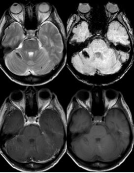
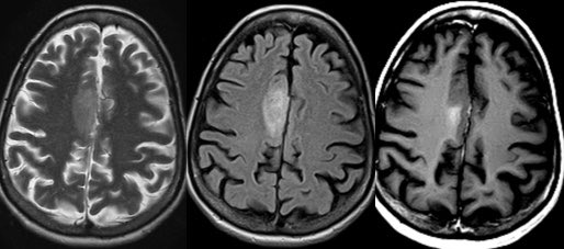
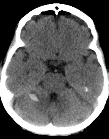
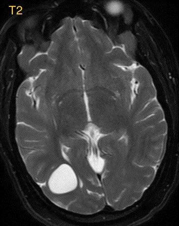
Answer: confirmed germinoma, all these masses are on the differential for a pineal region mass …perhaps the most helpful clue is the age and gender rather than the imaging 🧠
Germinoma cannot always be differentiated from pineoblastoma though the older age and male gender favor germinoma in this case as pineoblastoma typically occurs in younger children with a sight female predominance
Calcifications are also displaced and engulfed in this case rather than exploded or “blasted” and this tumor is very homogenous while pineoblastoma tends to be a bit more heterogenous
Meningioma arising from the tentorial cerebelli or falx can also look very similar though most commonly occur in older females around 5th-7th decades of life. Meningiomas tend to depress cerebral veins rather than uplift as pineal based masses do and may have a dural tail
There was no sellar lesion in this case, the mass effect caused the deformity of the pituitary stalk mimicking another lesion which resolved following resection

• • •

Missing some Tweet in this thread? You can try to force a refresh
 

Keep Current with Daniel Gewolb, MD

Daniel Gewolb, MD Profile picture

Stay in touch and get notified when new unrolls are available from this author!

Read all threads

This Thread may be Removed Anytime!

PDF

Twitter may remove this content at anytime! Save it as PDF for later use!

Try unrolling a thread yourself!

how to unroll video
  1. Follow @ThreadReaderApp to mention us!

  2. From a Twitter thread mention us with a keyword "unroll"
@threadreaderapp unroll

Practice here first or read more on our help page!

More from @daniel_gewolb

Mar 20
Glioblastoma is the most common variety of astrocytoma

The presence of necrosis is the characteristic feature of glioblastoma

Imaging details in thread #Neurosurgery #neurotwitter #radres #MedTwitter #Neurology @TheASNR
Some correlates for high grade astrocytoma include necrosis, marked mass effect, restricted diffusion (often patchy, eccentric or nodular in glioblastoma), elevated CBV, enhancement, and older age

The presence of ependymal spread is another poor prognostic sign (shown below)
Important learning point: when dealing with glioblastoma, the neoplasm extent is not defined by the area of enhancement but rather by the entire area of T2/FLAIR abnormality. Microscopic infiltrating tumor clearly extends beyond the areas of enhancement. 🧠
Read 5 tweets
Mar 16
Preoperative approach to sellar region masses, what the surgeon needs to know (at least what I think they need to know)

Additional reporting tips from surgeons are welcomed and encouraged! #Neurosurgery @TheASNR #radres #MedEd #MedTwitter #futureradres #endocrine #Neurology
1️⃣Where is the mass located?

Is it sellar based? suprasellar (S)? clival? Planum sphenoidale (PS)? Tuberculum sellae (arrow)?

▶️Location can change operative approach including but not limited to subfrontal vs transsphenoidal
2️⃣Where is the normal pituitary gland?

▶️this can be tough when the mass is large but the normal tissue often enhances more avidly than tumor so look for a strip of relatively avid enhancement along the periphery of the mass. Surgeons do not want to remove normal pit tissue
Read 10 tweets
Mar 14
Can you figure out the cause of hemorrhage in this case?

Imaging and case details in thread #Neurosurgery #radres #MedTwitter #Neurology @TheASNR #MedEd #neurotwitter ImageImage
Initial MRI shows an expansile enhancing mass in the right parasagittal frontal lobe ImageImageImageImage
The patient underwent craniotomy for tumor debulking. Post operative MRI and CT demonstrate hemorrhage in the right cerebellar hemisphere, far from the operative site. What’s the cause of the hemorrhage? 🤔 🧠 ImageImageImage
Read 5 tweets
Mar 6
What’s the most likely diagnosis in this 30 yr old presenting following an episode of shaking

More images in thread. Answer in a few hours 🧠 #radres #Neurosurgery #neurology #MedTwitter #neurotwitter @TheASNR Image
ImageImageImageImage
ImageImage
Read 6 tweets
Feb 26
Mesial Temporal Sclerosis (MTS) in this patient with temporal lobe seizures

Etiology: controversial ->
▶️possibly acquired from prolonged febrile seizures in infancy, perinatal ischemia, encephalitis, hypoxia from status epilepticus, etc.
▶️possibly developmental
#neurotwitter Image
Etiology: probably best thought of as a common outcome of multiple acquired and developmental processes

▶️The hippocampus is incredibly sensitive to anoxia (in particular Ammon’s horn)
▶️Ammon’s horn has 4 zones of granular cells
▶️zone CA1 is the most sensitive area of the brain to anoxia and as such, is the main location for the disease

Imaging findings:
T1-> diminished size and loss of GW diff
Quantitative hippocampal volumes can improve sensitivity especially in the cases of bilateral disease #radres
Read 6 tweets

Did Thread Reader help you today?

Support us! We are indie developers!


This site is made by just two indie developers on a laptop doing marketing, support and development! Read more about the story.

Become a Premium Member ($3/month or $30/year) and get exclusive features!

Become Premium

Don't want to be a Premium member but still want to support us?

Make a small donation by buying us coffee ($5) or help with server cost ($10)

Donate via Paypal

Or Donate anonymously using crypto!

Ethereum

0xfe58350B80634f60Fa6Dc149a72b4DFbc17D341E copy

Bitcoin

3ATGMxNzCUFzxpMCHL5sWSt4DVtS8UqXpi copy

Thank you for your support!

Follow Us on Twitter!

:(