I'm excited to share my latest paper in @JASN_News on #complementbiology:
"Non-Invasive Detection of iC3b/C3d Deposits in the Kidney Using a Novel Bioluminescent
Imaging Probe."
pubmed.ncbi.nlm.nih.gov/36995143/
Time for a 🧵 & #tweetorial 👇
"Non-Invasive Detection of iC3b/C3d Deposits in the Kidney Using a Novel Bioluminescent
Imaging Probe."
pubmed.ncbi.nlm.nih.gov/36995143/
Time for a 🧵 & #tweetorial 👇

🧵(1/10): Histologic quantification of complement C3 deposits in kidney biopsies provides prognostic information in patients with:
- IgA nephropathy,
- ANCA-associated glomerulonephritis,
- Membranous nephropathy,
- Focal segmental glomerulosclerosis,
- Diabetic nephropathy.
- IgA nephropathy,
- ANCA-associated glomerulonephritis,
- Membranous nephropathy,
- Focal segmental glomerulosclerosis,
- Diabetic nephropathy.
🧵(2/10): As patients are not biopsied repeatedly (for obvious reasons!), histological examination only offers a single “snapshot” of a portion of a kidney at the time of sampling, which may not accurately represent the ongoing pathological process in both kidneys over time.
🧵(3/10): Molecular imaging for C3 deposition offers the promise of non-invasively detecting complement activation in live animals. A challenge in detecting C3 deposits is that intact C3 is abundant in🩸. The probe must thus distinguish tissue-bound C3 fragments from C3 in🩸.
🧵(4/10): My mentors, Prof. Thurman & Holers @CUAnschutz, previously developed a panel of anti-human C3d mAbs @jclinicalinvest, that cross-reacts with mouse C3d, target tissue-bound C3d while not binding intact C3, making them an ideal probe for imaging⬇️
pubmed.ncbi.nlm.nih.gov/23619360/
pubmed.ncbi.nlm.nih.gov/23619360/
🧵(5/10): In this study, we developed a method to non-invasively detect C3 fragment deposition throughout both kidneys, using the 3d29 monoclonal antibody targeting tissue-bound C3d linked to a bioluminescent resonance energy transfer construct that emits near-infrared light.
🧵(6/10): We used a reporter construct that contains a NanoLuc luciferase (nLUC) & 2️⃣red-shifted orange fluorescent proteins (OFPs). When the coelenterazine substrate reacts with the nLUC (step 1), this emits light (step 2), which excites the OFPs to emit light at 590nm (step 3). 

🧵(7/10): The probe will thus emit light of near-infrared, which gives very little background signal, and penetrate tissues to a depth of 5 mm to 3 cm.
Although this range is not sufficient for imaging deep tissues in humans, it can be used to detect molecular targets in mice.
Although this range is not sufficient for imaging deep tissues in humans, it can be used to detect molecular targets in mice.
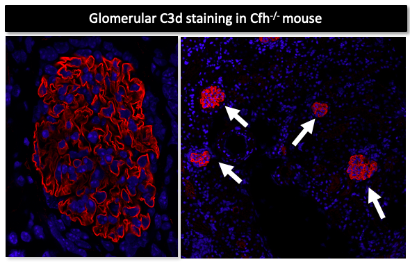
🧵(8/10): We tested whether the reporter-conjugated anti-C3d mAb could be used to detect renal C3d deposition in a mouse model of C3 glomerulopathy (Cfh-/- mice).
These mice have abundant glomerular deposits of C3d when you perform immunohistochemistry on their renal sections ⬇️
These mice have abundant glomerular deposits of C3d when you perform immunohistochemistry on their renal sections ⬇️

🧵(9/10): When we performed imaging in live animals, the kidneys of Cfh-/- mice emitted strong signal in vivo compared to WT & C3-/- control mice (image⬇️)
The kidneys of Cfh-/- mice injected with a control mAb or untargeted reporter emitted very little signal (data not shown).
The kidneys of Cfh-/- mice injected with a control mAb or untargeted reporter emitted very little signal (data not shown).

🧵(10/10): In summary, we developed a method to non-invasively detect & quantify C3d deposits throughout both kidneys of live mice.
Biopsies look at a small region of the kidney, but molecular imaging may more accurately assess the overall degree of inflammation in both kidney.
Biopsies look at a small region of the kidney, but molecular imaging may more accurately assess the overall degree of inflammation in both kidney.
• • •
Missing some Tweet in this thread? You can try to
force a refresh


