Ritu Thamman MD Profile picture
Associate Professor #cardiologist #SoMe editor @CircOutcomes @JournalASEcho #BOD @ase360 @PittHealthSci @ColumbiaMed @NUFeinbergMed #echofirst #NewJersey girl
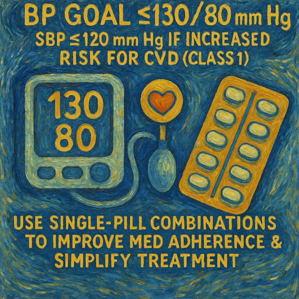
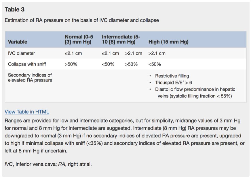
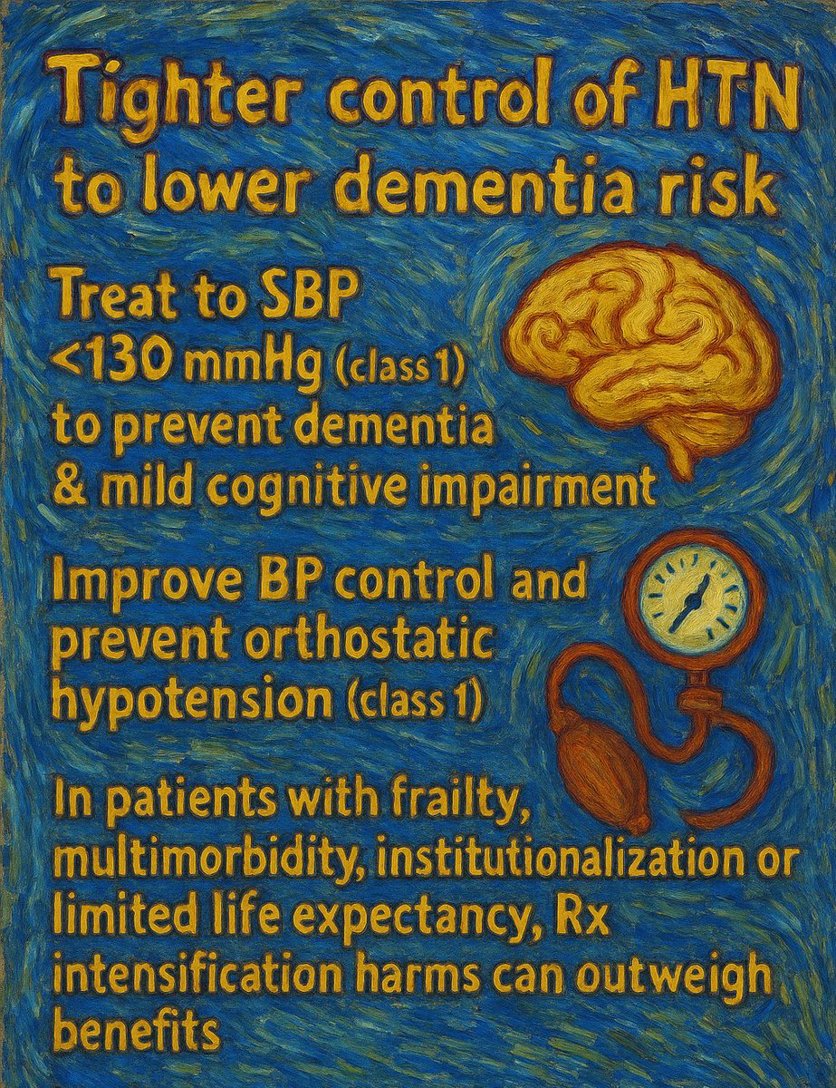
Karen Zimmerman Profile picture Reza Hosseini, MD MPH Profile picture Simone Ferreira Leite Profile picture Juan Jose Rousseau Profile picture 5 subscribed
Mar 2, 2021 10 tweets 12 min read
Tweetorial on Challenges in Quantification of Aortic stenosis before tonight’s #ASEchoJC on @PPibarot & @E_Guzzetti 📝 bit.ly/2NNIJgC

~1/3 pts have DISCORDANT indices: AVA is severe <1 cm2 yet mean gradient is low <40 mmHG bit.ly/3dWmJuy

Low Gradient types👇 2/low gradient severe AS
types:

1.Classic:both flow SVI EF⬇️(classical Low flow CLF)

2. Paradoxical: EF nl BUT low flow SVI (Paradoxical Low flow PLF )

3. Both EF and flow are nl (Normal Flow NF)

Low flow⬆️💀mortality
Paradoxical Low flow &
Classical Low flow #ASEchoJC
Oct 27, 2020 8 tweets 6 min read
@hahn_rt 📝 #ASEchoJC

Can a single cycle length method can be used to calculate aortic EOA in aortic stenosis w nonsinus rhythms?

100 AS pts w R-R variability identified:55 w atrial fib AF & 45 w frequent atrial or ventricular premature contractions FE bit.ly/3or3ufk 2/#ASEchoJC
LVOT TVI by PWD & AV VTI CWD measured over 5-10 consecutive beats in AF group & over 3-5 consecutive sinus beats in FE,EOA & DVI calculated as guidelines standard
In all patients, LVOT diameter was measured in midsystole, within 2-4 mm apical to annulus
Sep 13, 2020 10 tweets 10 min read
#ASEchoJC #Tweetorial on #WASE
World
Alliance
Societies of Echocardiography
Normal Values Study

#Echofirst most widely used imaging modality BUT

WHAT IS NORMAL for ❤️ size & function?

bit.ly/32pKxAC @JournalASEcho 2/ Prior ❤️measurement studies:
❤️used VARIOUS methods- M mode, M mode & simpsons, 2D
❤️No standard analysis or core lab
❤️Only single race or country/region
❤️?? State of the art machines or techniques

❤️Need for a prospective international observational study #WASE was born
Jan 22, 2019 16 tweets 9 min read
1/#Tweetorial 3D Echo(TTE/TEE) & CT methods Tricuspid annulus & valve assessment in severe TR #ASEechoJC Today 8 pm
bit.ly/2FFWk3I
pts with severe tricuspid regurgi, semiautomated indirect planimetry results in agreement b/w TEE & CT for Tricuspid annulus sizing & TVA 
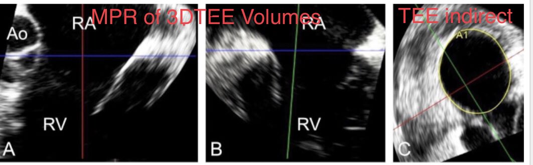
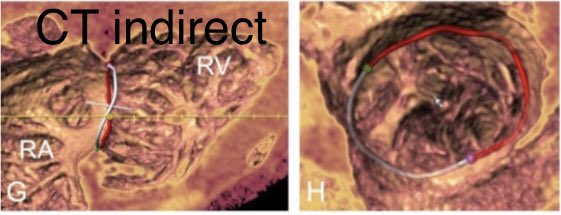
2/TEE_direct of the TA allows the most accurate measurement of diastolic stroke volume for the calculation of regurgitation severity compared with 3D vena contracta area.

This is first study to validate use of 3D TEE planimetered annular area to quantify the regurgitant volume
Dec 8, 2018 10 tweets 18 min read
1/Tweetorial on #TMVR for #ASEchoJC 12/11 8 pm
bit.ly/2EiJQ1v

transcatheter mitral valve replacement #TMVR for failed valve & ⬆️ #STS risk

#VHD 2017 update reoperation➡️ reintervention bit.ly/2P9bX4G

🎈expandable SAPIEN S3 only @FDA bioprosthesis for implant 2/#Transeptal developed @MayoClinicCV 48 pts

After 🇨🇦 initial experience with #transapical 2009

sick patients with ⬇️ EF #transeptal preserves EF better avoids apical purse string suture

Currently #TMVR registry
>50 % are transeptal & in house mortality 7%
Oct 22, 2018 10 tweets 18 min read
1/#Tweetorial #papillary #fibroelastoma for
#ASEchoJC 10/23

🐙= #PFE 1975 named #papillary #fibroelastoma by #armed #forces #institute of #pathology

#papilloma 1973

#Collagen core with elastic fibers & matrix covered by endothelium sheath
Attached by stalk
multiple fronds 
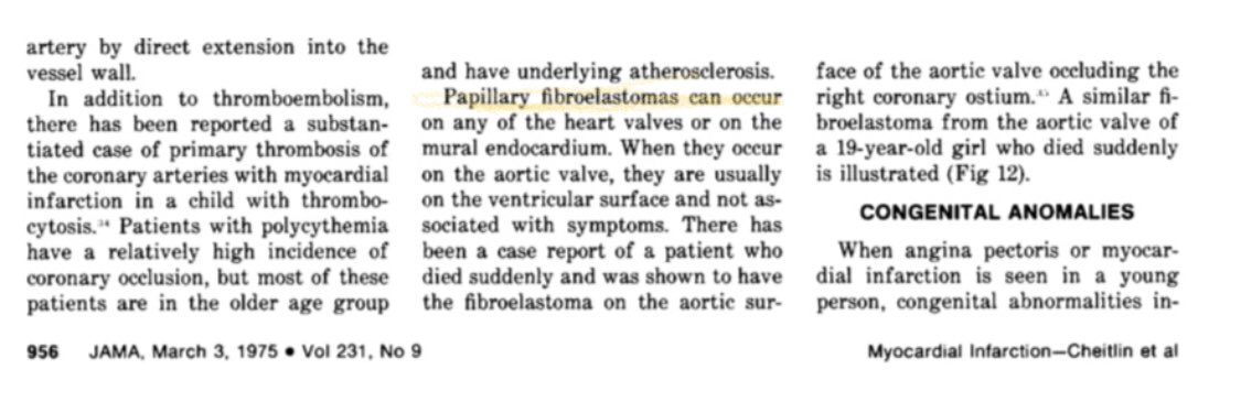
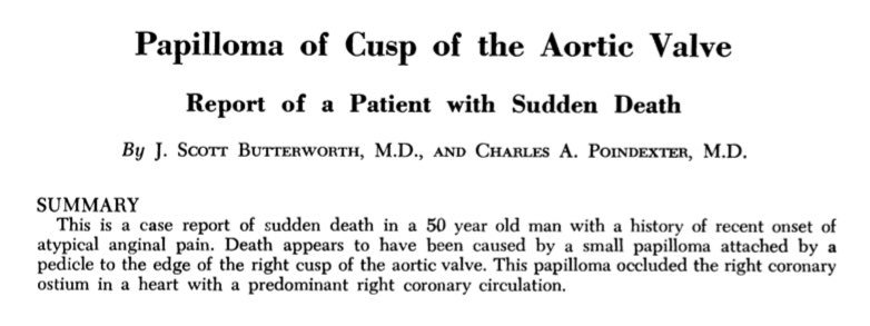
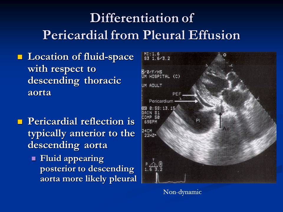
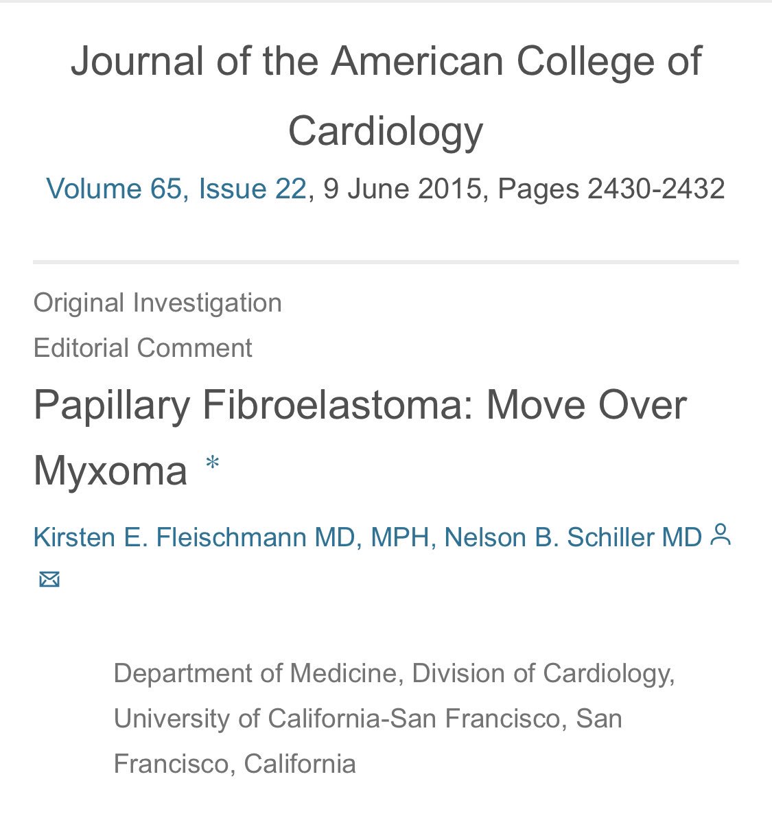
2/ What is the incidence of #PFE 🐙 compared to #myxoma (diagnosed in same time period)?

#PFE 511 vs. #myxoma 112

#PFE incidence ~1/1000

common “benign”primary ❤️tumor #modern #EchoFirst (Most common @MayoClinicCV)

#myxoma (autopsy based prevalence)

bit.ly/2Am05Y9
Sep 3, 2018 10 tweets 16 min read
Curious about how #POCUS is taught? This #Tweetorial is for U
#Preview for #ASEchoJC 🔜9/4 8pm EST

Point-of-Care Cardiac Ultrasound POCUS: State-of-the-Art in Medical School Education bit.ly/2wxD4PZ by @amerjohri

#POCUS not 🐇🎩, not short #TTE 
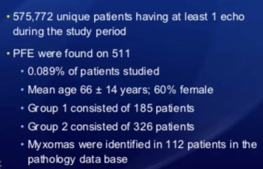
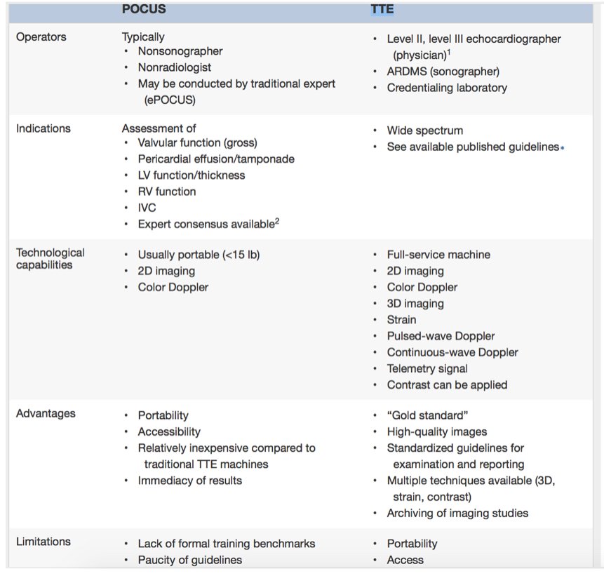
2/ Current #goals #Cardiac #POCUS #MedEd

(1) introduce concepts of ultrasound- common imaging views, correlate with anatomy, & physical examination skills

(2) develop scanning techniques➡️ basic competence

(3) recognize & differentiate b/w normal anatomy & basic pathology