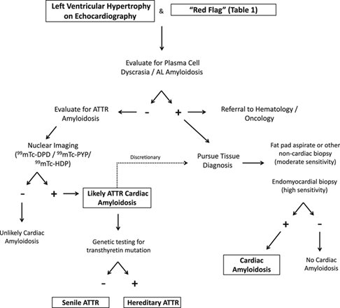
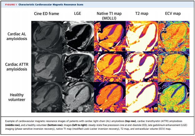
As #ESCCongress nears, I thought I would do a #tweetorial on amyloidosis. Exciting times for the field and new data/treatments expected next week.
#FITSurvivalGuide #CardioTwitter @tony_breu @rodney_falk @marthagrogan1 @amyloidosisfdn @AmyloidosisSupp @Amyloidosis_ARC
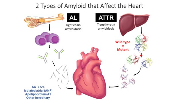
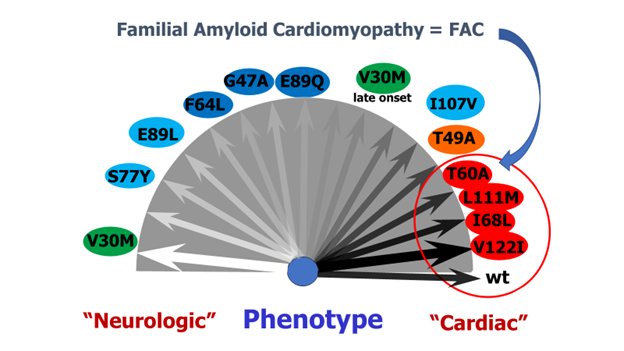
ATTR amyloidosis may occur as a result of the wild-type protein or from a hereditary mutation. This fantastic figure from Rapezzi et al depicts the common mutations on a spectrum from cardiomyopathy to neuropathy.
ncbi.nlm.nih.gov/pubmed/22745357
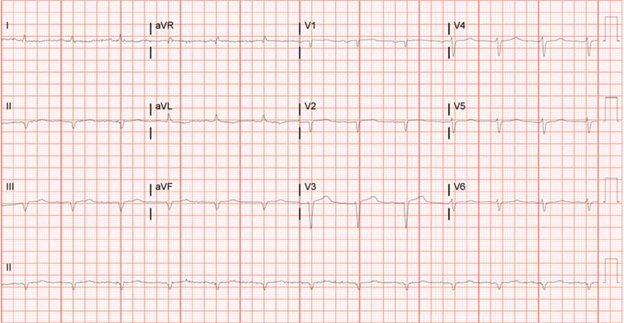
ECG is *sometimes* low voltage and/or pseudoinfarct pattern. ECG voltage to echo mass ratio is more often abnormal which should set off alarms to consider amyloidosis.
ncbi.nlm.nih.gov/pubmed/27093686
ncbi.nlm.nih.gov/pubmed/25212550
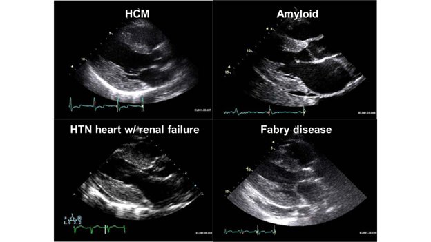
Typical echo characteristics are increased LV wall thickness and diastolic dysfunction. Sometimes valve and IAS thickening. “Sparkling” myocardium not specific with harmonic imaging. Do you see the decreased longitudinal motion on the 2d images?
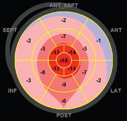
We know strain is helpful to reclassify patients with “LVH”, particularly into the amyloidosis category.
ncbi.nlm.nih.gov/pubmed/24874973
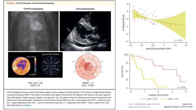
Look at the SPECT images as well. You may see an apical sparing pattern (ie less diffuse deposition) which portends a better prognosis.
ncbi.nlm.nih.gov/pubmed/28917675
