Neuroanatomy TOTD #4
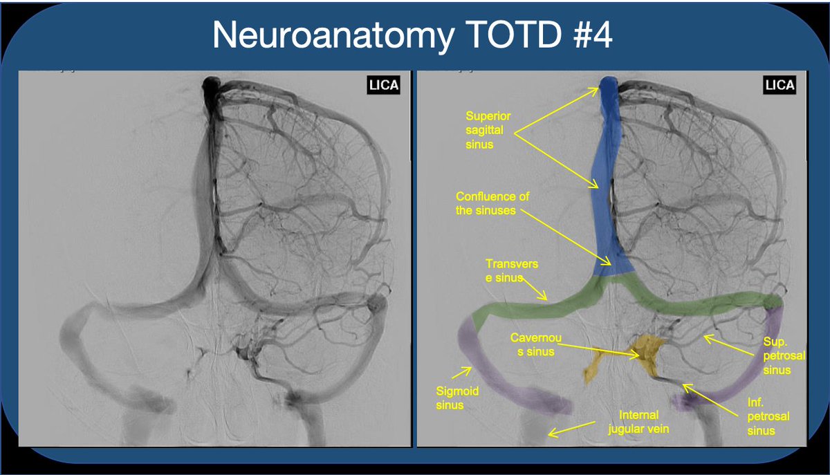
1/5 Answer: The orange structure is the cavernous sinus (CS), a paired dura-lined venous cavity on either side of the sella. The sinuses are split into numerous “caves” by fibrous septae (hence the name). #neuroanatomy #neurorad #medtwitter #neuroanatomyTOTD Image
2/5 Note that the paired sinuses are often variably connected by “intercavernous sinuses”. While the CS is often taught for its relationship to the ICA and cranial nerves, I find that medical students and residents rarely understand the flow of blood through the sinus.
3/5 The CS receives blood from sup. and inf. ophthalmic veins draining the orbit--This is how a facial/orbital infection spreads intracranially to CS (classic #usmle ?). The sphenoparietal sinus as well as the superficial middle and inferieor cerebral veins also feed into the CS. Image
4/5 The CS drains via the sup & inf petrosal sinuses (to the transverse/sigmoid sinus & sigmoid/int jug), as well as variable drainage through emissary veins which course through skull foramina (like the foramen ovale). This includes variable connections to the pterygoid plexus. Image
5/5 Understanding crucial for interventional closure of carotid-cavernous AV fistula. Can be approached: retrograde via IJV/inf petrosal sinus, anterograde/transorbital through sup ophth vein, +/- arterial approach via ICA. #meded #FOAMed #FOAMrad #radres #radiology #neurosurgery Image

• • •

Missing some Tweet in this thread? You can try to force a refresh
 

Keep Current with Aaron Rutman, MD

Aaron Rutman, MD Profile picture

Stay in touch and get notified when new unrolls are available from this author!

Read all threads

This Thread may be Removed Anytime!

PDF

Twitter may remove this content at anytime! Save it as PDF for later use!

Try unrolling a thread yourself!

how to unroll video
  1. Follow @ThreadReaderApp to mention us!

  2. From a Twitter thread mention us with a keyword "unroll"
@threadreaderapp unroll

Practice here first or read more on our help page!

More from @aaronrutman

30 Oct
Neuroanatomy TOTD #10🧵 
1/5 Small gray matter structure at the junction of the thalamus and midbrain is the subthalamic nucleus (STN).

#meded #FOAMed #FOAMrad #medtwitter #medstudents #radiology #neurorad #radres #neurology #neurosurgery #neuroanatomy #neuroanatomyTOTD Image
2/5...The STN is functionally a node within the basal ganglia (BG) INDIRECT LOOP. STN contains excitatory glutaminergic neurons➞output to the GABA neurons of GPi, which in turn have inhibitory effect on thalamic outputs to the motor cortex. Image
3/5...Loss of nigrostriatal input in Parkinsons Dz➞increased inhibitory output from GPi➞decreased thalamic stimulation of the motor cortex (through both direct and indirect loop circuits). Makes sense that DBS treatments were initially directed at disrupting activity in GPi. Image
Read 5 tweets
29 Sep
Neuroanatomy TOTD #6
a) Ant choroidal artery infarct, involving the post limb of the internal capsule
b) Sup hypophyseal artery aneurysm—medially directed from the supraclinoid ICA
#meded #FOAMed #FOAMrad #radres #neurorad #radiology #neurosurgery #neuroanatomy #neuroanatomyTOTD ImageImage
ICA segments: C1 (cervical) becomes C2 (petrous) in the carotid canal of the petrous bone. Becomes C3 (lacerum) as it exits the carotid canal above the foramen lacerum. Becomes C4 (cavernous segment above the petrolingual ligament through the cavernous sinus. Image
...C5 (clinoid) above proximal to the distal dural ring. C6 (ophthalmic) is truly intracranial. C7 (communicating) distal to the Pcomm. Alternative segmentation schemes include C1-C4 (cervical, petrous, cavernous, supraclinoid/terminal) Image
Read 5 tweets
24 Sep
Neuroanatomy TOTD #5

The indicated bundle is the anterior commissure (AC), located at the ant border of the 3rd ventricle, at the sup margin of the lamina terminalis.

#meded #FOAMed #FOAMrad #radres #neurorad #medtwitter #radiology #neurosurgery #neuroanatomy #neuroanatomyTOTD Image
The AC runs across the midline in front of the anterior columns of the fornix, behind the basal forebrain and beneath the anterior limb internal capsule and basal ganglia, surrounded by the bed nucleus of the stria terminalis.
The AC connects areas of the bilateral temporal poles and orbitofrontal cortex. Function is not entirely understood but it is thought to be important in the olfactory pathway and pain sensation, among other things.
Read 6 tweets
19 Sep
Neuroanatomy TOTD #3
1/6 The superior frontal sulcus courses in the AP direction, and terminates at the precentral sulcus posteriorly. The central sulcus is immediately posterior to the precentral, and the postcentral immediately posterior to the central.
#meded #FOAMed #FOAMrad Image
2/6 "Upper T sign" or "L sign"
The SFS intersects the precentral s
Central sulcus is just posterior Image
3/6 "Lower T sign" or "M sign"
The IFS terminates at the precentral s.
Central sulcus is just posterior Image
Read 7 tweets
19 Sep
Neuroanatomy TOTD #2:

Answer: Fetal PCA

1/3 A fetal PCA, or fetal origin of the PCA is a common variant, estimated to be present (at least on one side) in about a quarter of individuals. Image
2/3 The term is used variably, when the bulk of PCA flow comes from the anterior circulation (internal carotids) via a robust post. communicating artery. Some reserve the term for when there is no connection to the posterior circulation (absent P1 segment from the basilar art.), Image
3/3 ...while others use the term any time the Pcomm is the dominant supply to the PCA, (rather than the basilar and P1 segment). Note that when fetal PCA is bilateral, the vertebral and basilar arteries are usually diminutive, terminating in superior cerebellar arteries. Image
Read 4 tweets

Did Thread Reader help you today?

Support us! We are indie developers!


This site is made by just two indie developers on a laptop doing marketing, support and development! Read more about the story.

Become a Premium Member ($3/month or $30/year) and get exclusive features!

Become Premium

Too expensive? Make a small donation by buying us coffee ($5) or help with server cost ($10)

Donate via Paypal Become our Patreon

Thank you for your support!

Follow Us on Twitter!