Want to figure out why your 🚑🩸Trauma Patient is so Hypotensive? Master the #POCUS eFAST Exam TODAY!
1⃣Learn the eFAST Exam in 5 Easy Steps
2⃣Abdominal/Thoracic Free Fluid
3⃣Cardiac Tamponade
4⃣Pneumothorax
✅New Blog Post
🔗👉pocus101.com/eFAST
#medtweetorial 👇(1/23)
1⃣Learn the eFAST Exam in 5 Easy Steps
2⃣Abdominal/Thoracic Free Fluid
3⃣Cardiac Tamponade
4⃣Pneumothorax
✅New Blog Post
🔗👉pocus101.com/eFAST
#medtweetorial 👇(1/23)


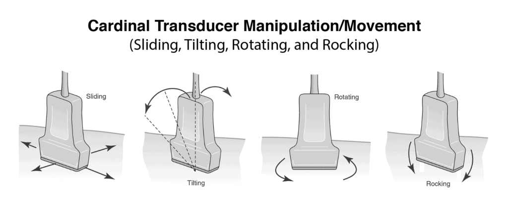
(2/) Using a Phased Array or Curvilinear probe. Position the patient supine (or Trendelenburg).
🔗👉pocus101.com/eFAST
🔗👉pocus101.com/eFAST

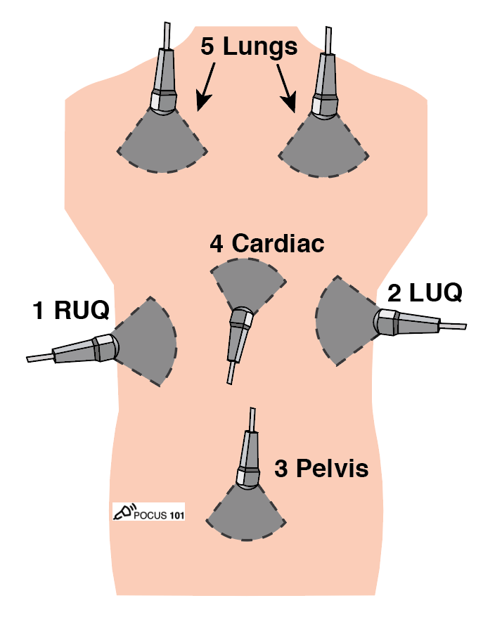
(3/) Here are the 5 steps we recommend for the eFAST:
1 Right Upper Quadrant View (RUQ)
2 Left Upper Quadrant View (LUQ)
3 Pelvic View
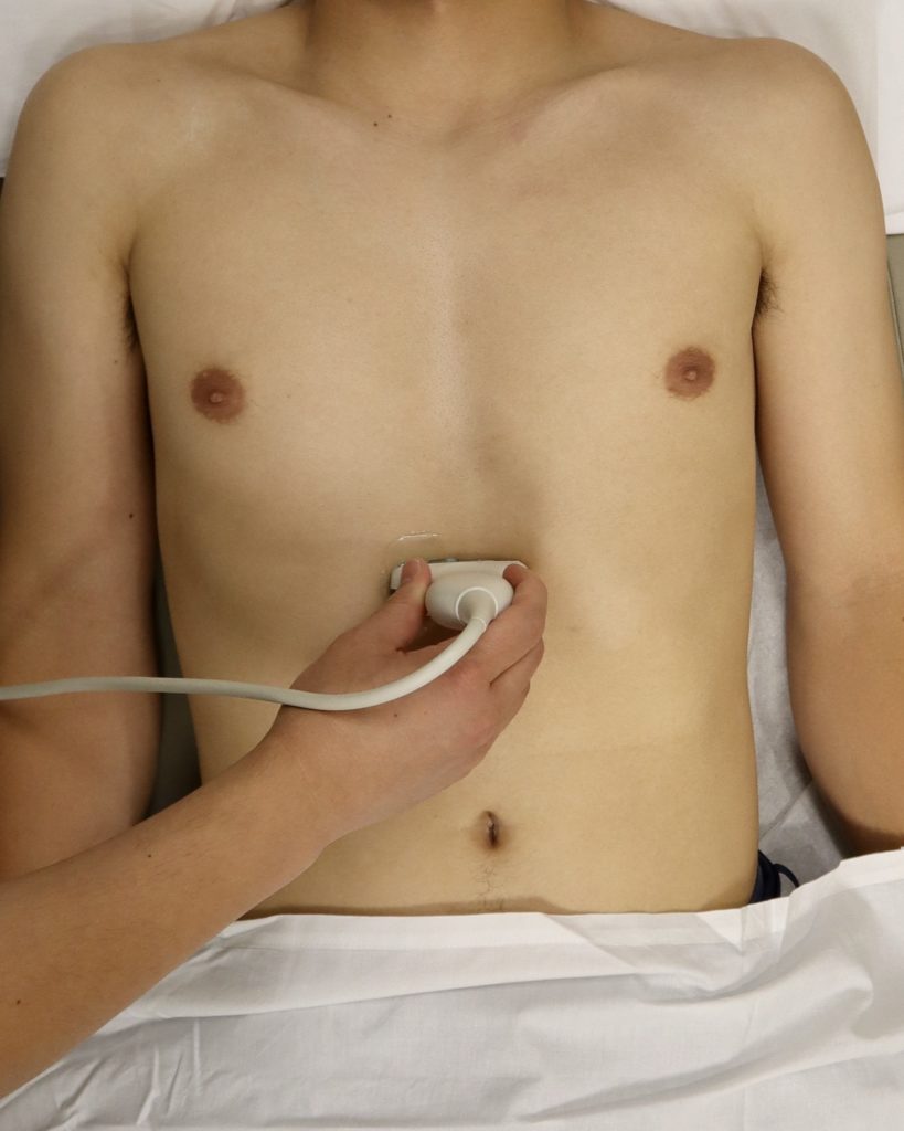
4 Cardiac View (Parasternal Long Axis or Subxiphoid)
5 Lungs (Right and Left)
🔗👉pocus101.com/eFAST
1 Right Upper Quadrant View (RUQ)
2 Left Upper Quadrant View (LUQ)
3 Pelvic View
4 Cardiac View (Parasternal Long Axis or Subxiphoid)
5 Lungs (Right and Left)
🔗👉pocus101.com/eFAST
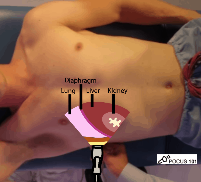
(4/) RUQ View
Orientate the probe indicator towards the patient’s head.
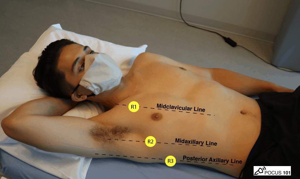
Anchor your probe in the midaxillary line at the 10th intercostal space.
🔗👉pocus101.com/eFAST
Orientate the probe indicator towards the patient’s head.
Anchor your probe in the midaxillary line at the 10th intercostal space.
🔗👉pocus101.com/eFAST


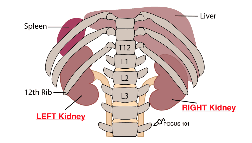
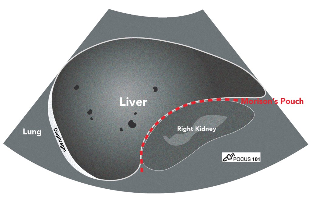
(5/) Using the liver as an acoustic window, identify the lung, liver, Morison’s Pouch, diaphragm, and the long-axis of the right kidney.
🔗👉pocus101.com/eFAST
🔗👉pocus101.com/eFAST

(6/) Here is what an Abnormal RUQ view can look like with fluid in Morison's Pouch and Caudal tip of the Liver.
🔗👉pocus101.com/eFAST
🔗👉pocus101.com/eFAST
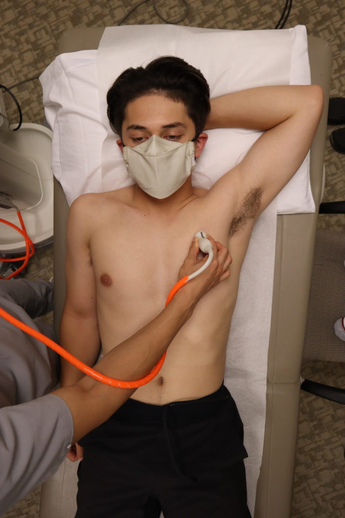
(7/) LUQ View
Orientate the probe indicator towards the patient’s head.
Anchor your probe in the posterior axillary line around the 8th intercostal space.
You should have your “Knuckles to the bed” since the spleen is fairly posterior.
🔗👉pocus101.com/eFAST
Orientate the probe indicator towards the patient’s head.
Anchor your probe in the posterior axillary line around the 8th intercostal space.
You should have your “Knuckles to the bed” since the spleen is fairly posterior.
🔗👉pocus101.com/eFAST


(8/) Using the spleen as an acoustic window, identify the spleen, perisplenic space, diaphragm, and the long-axis view of the left kidney.
🔗👉pocus101.com/eFAST
🔗👉pocus101.com/eFAST


(9/) Here is what an Abnormal LUQ view can look like with fluid in the Perisplenic Space (between diaphragm and spleen).
🔗👉pocus101.com/eFAST
🔗👉pocus101.com/eFAST
(10/) Don't forget to look above the diaphragm in the RUQ and LUQ views to rule out Hemothorax with the Spine Sign!
🔗👉pocus101.com/eFAST
🔗👉pocus101.com/eFAST
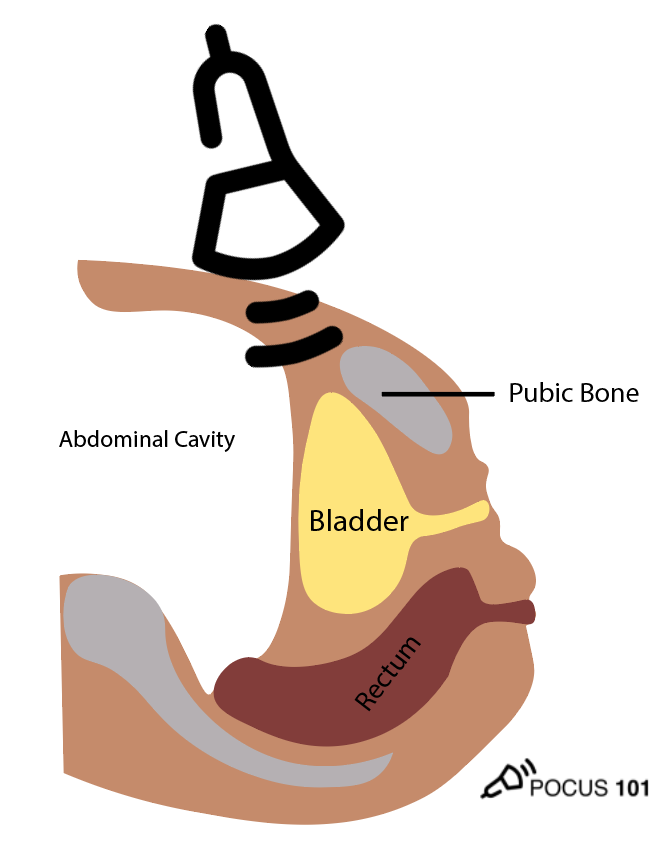
(11/) Pelvic Longitudinal View
Place the transducer with the indicator pointing towards the patient’s head in the patient’s midline, right above the pubic symphysis.
Rock the probe so that it points down towards the pelvic cavity.
🔗👉pocus101.com/eFAST
Place the transducer with the indicator pointing towards the patient’s head in the patient’s midline, right above the pubic symphysis.
Rock the probe so that it points down towards the pelvic cavity.
🔗👉pocus101.com/eFAST


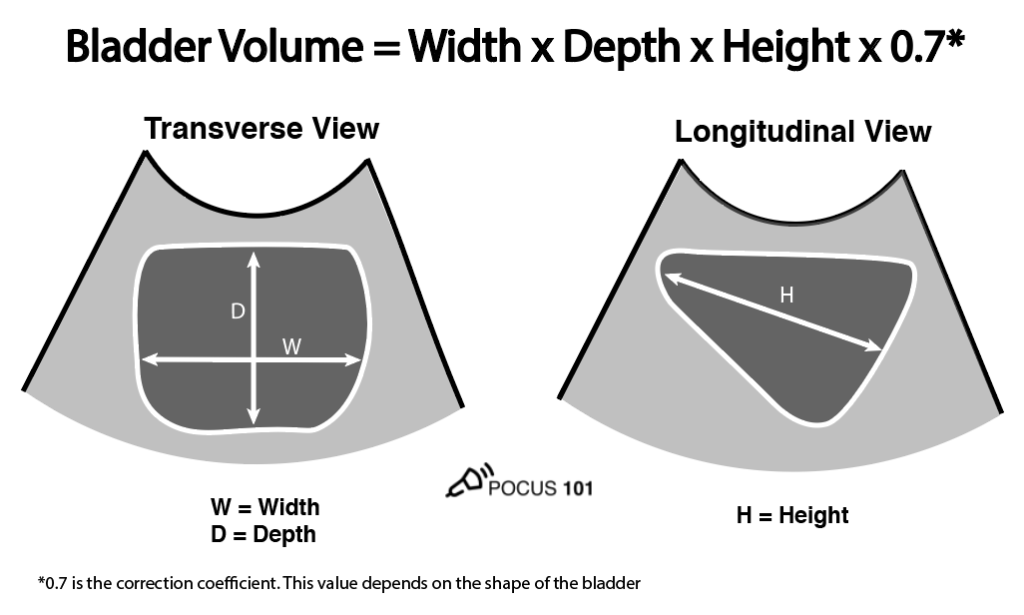
(12/) Pelvic Transverse View
Center the bladder and then rotate the transducer 90 degrees counterclockwise. The indicator should now point to the patient’s Right side.
Make sure to tilt the ultrasound probe so it scans into the pelvic cavity.
🔗👉pocus101.com/eFAST
Center the bladder and then rotate the transducer 90 degrees counterclockwise. The indicator should now point to the patient’s Right side.
Make sure to tilt the ultrasound probe so it scans into the pelvic cavity.
🔗👉pocus101.com/eFAST

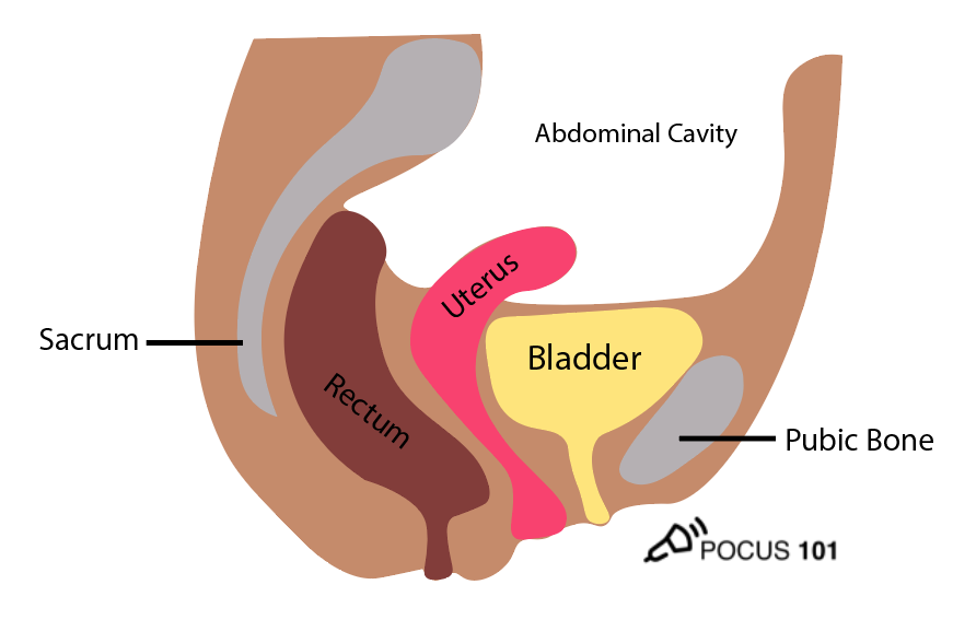
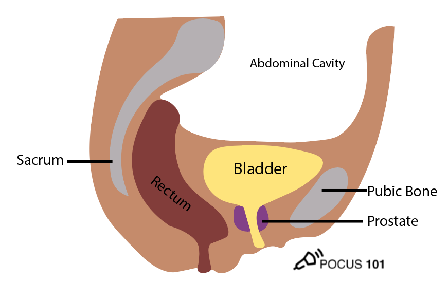
(13/) For Males look for free fluid in the Rectovesical Pouch and for Females look for free fluid in the Pouch of Douglas.
🔗👉pocus101.com/eFAST
🔗👉pocus101.com/eFAST


(16/) Cardiac Views
Use either the subxiphoid view or the parasternal long axis views to rule out pericardial effusion.
Here is an example of a normal subxiphoid view.
🔗👉pocus101.com/eFAST
Use either the subxiphoid view or the parasternal long axis views to rule out pericardial effusion.
Here is an example of a normal subxiphoid view.
🔗👉pocus101.com/eFAST
(18/) To diagnose tamponade, look for either right atrial systolic collapse or right ventricular diastolic collapse.
🔗👉pocus101.com/eFAST
🔗👉pocus101.com/eFAST
(19/) Lastly, scan the right and left lung to rule out Pneumothorax. Place the probe in the mid-clavicular line around the 2nd intercostal space.
🔗👉pocus101.com/eFAST
🔗👉pocus101.com/eFAST

(20/) If lung sliding is present you have effectively ruled OUT pneumothorax! Easy Peasy.
🔗👉pocus101.com/eFAST
🔗👉pocus101.com/eFAST
(21/) If lung sliding is absent consider looking for the Lung Point Sign to confirm there is a pneumothorax.
🔗👉pocus101.com/eFAST
🔗👉pocus101.com/eFAST
(22/) Pitfalls/Limitations of the eFAST Exam:
1⃣Does not localize the injured abdominal organ
2⃣Limited views in patients with subcutaneous emphysema
3⃣Views may be limited in patients with hollow-viscus injury + free air in the abdomen (see image)
🔗👉pocus101.com/eFAST
1⃣Does not localize the injured abdominal organ
2⃣Limited views in patients with subcutaneous emphysema
3⃣Views may be limited in patients with hollow-viscus injury + free air in the abdomen (see image)
🔗👉pocus101.com/eFAST

(23/) Here is a simple eFAST algorithm you can use when scanning.
Remember the eFAST exam is most beneficial in unstable trauma patients who can't go to the CT scanner.
🔗👉pocus101.com/eFAST
Remember the eFAST exam is most beneficial in unstable trauma patients who can't go to the CT scanner.
🔗👉pocus101.com/eFAST

• • •
Missing some Tweet in this thread? You can try to
force a refresh