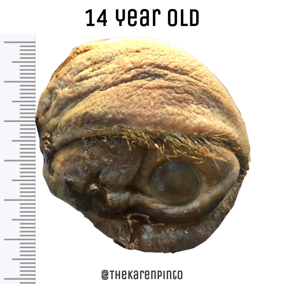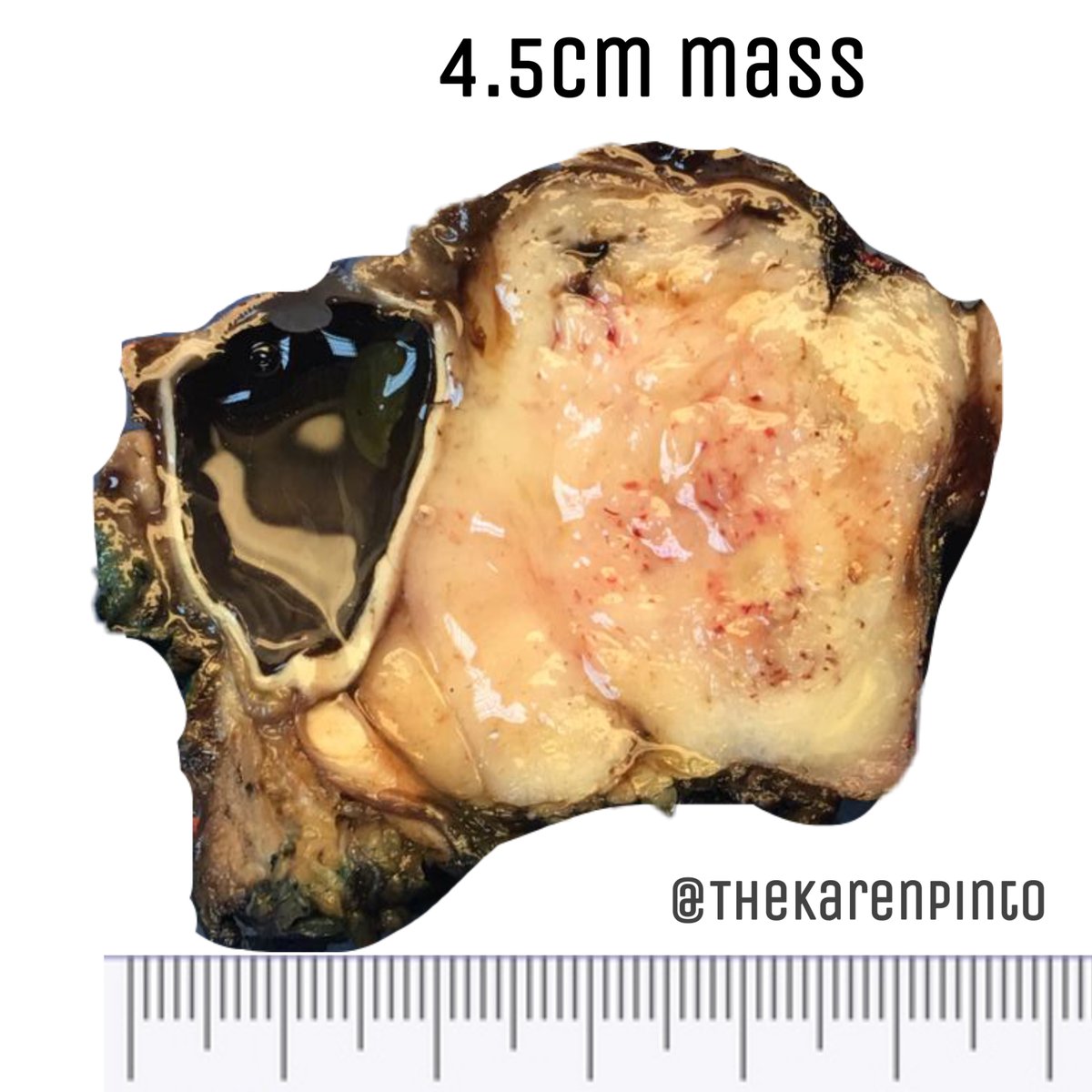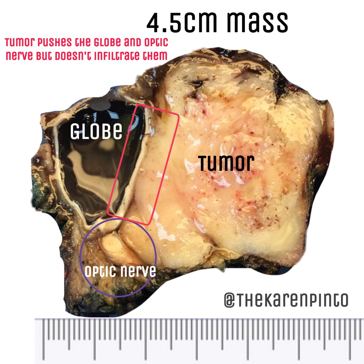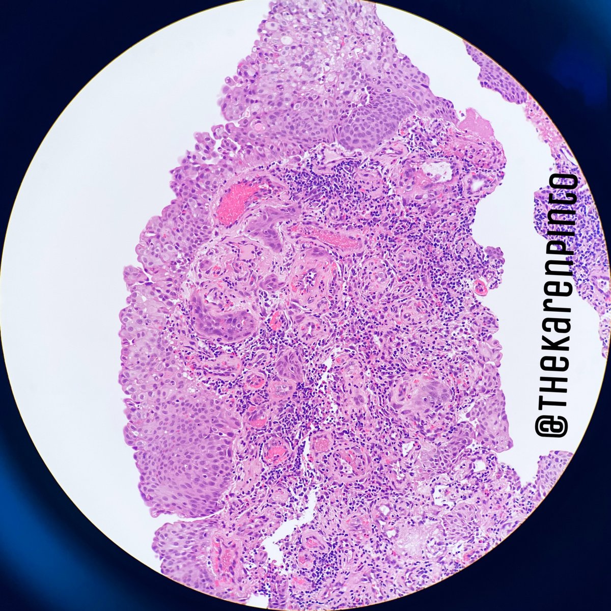
All you need to know about grossing the #larynx
Here's a #PathVideoTweetorial
#pathology #grosspath #headandneckpath
Video 1: Let's first get oriented by seeing some images
Here's a #PathVideoTweetorial
#pathology #grosspath #headandneckpath
Video 1: Let's first get oriented by seeing some images
Since the main tumor completely involved the epiglottis, the surgeon had to excise a part of the tongue (which is seen in the specimen)
Now that you’ve seen the images, let’s take a look at the real deal
Video 2: Larynx specimen intact
Now that you’ve seen the images, let’s take a look at the real deal
Video 2: Larynx specimen intact
After I took the cut margins, I had to bisect the specimen
(This is how I gross the specimen, others may have their own techniques)
Video 3: Bisecting the specimen
(This is how I gross the specimen, others may have their own techniques)
Video 3: Bisecting the specimen
To make it easier to understand, I've taken images and explained them
Video 4: Images of the specimen after cutting open the posterior half
Video 4: Images of the specimen after cutting open the posterior half
Video 5: What are the different parts of the larynx (as you see it in the actual specimen)
Video 6: Once the specimen has been bisected, we need to assess the extent of the tumor
Here it important to see it’s relation to the pre-epiglottic fat (involvement of the latter, upstages the tumor)
Here it important to see it’s relation to the pre-epiglottic fat (involvement of the latter, upstages the tumor)
Another important landmark is the pyriform fossa
The mucosa lining the fossa may not always be removed entirely (since it’s used for flap closure)
So you need to be aware what the surgeon has taken out
Video 7: Pyriform fossa
The mucosa lining the fossa may not always be removed entirely (since it’s used for flap closure)
So you need to be aware what the surgeon has taken out
Video 7: Pyriform fossa
The next step is to serially slice each half of the larynx and submit sections to show the extent of involvement of the tumor
Sometimes the cartilages are extensively calcified, making it difficult to cut
Video 8: Serial sectioning
Sometimes the cartilages are extensively calcified, making it difficult to cut
Video 8: Serial sectioning
Last but not the least, what is the paraglottic space?
Video 9: Paraglottic space and how to recognize it
Video 9: Paraglottic space and how to recognize it
I hope these videos help understand the anatomy and how to gross a specimen of the larynx better
I would like to acknowledge my amazing tech @Nora_almuttairi for recording all the above videos
& my awesome surgeon Dr. Hany Eldweny who I’ve learnt so much from
✌🏽
I would like to acknowledge my amazing tech @Nora_almuttairi for recording all the above videos
& my awesome surgeon Dr. Hany Eldweny who I’ve learnt so much from
✌🏽
• • •
Missing some Tweet in this thread? You can try to
force a refresh

