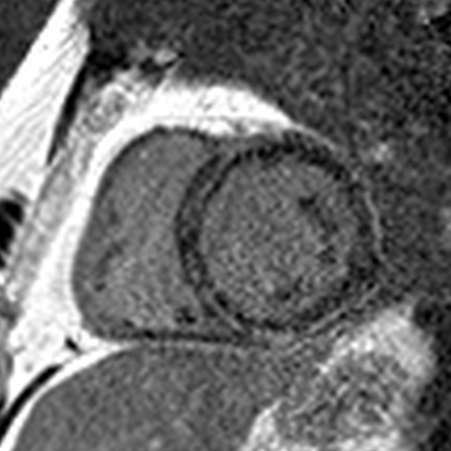
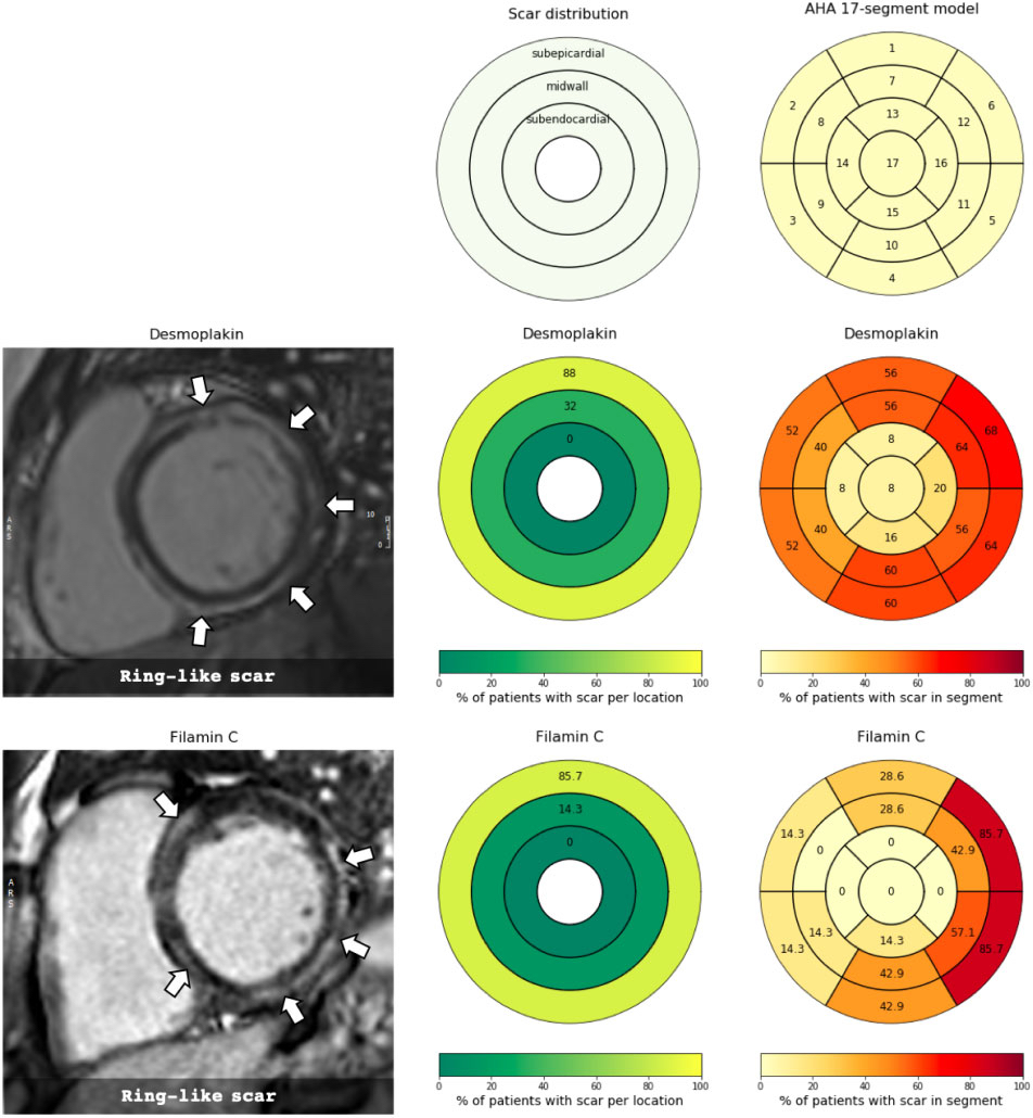
I think it’s genetic ACM because of the pattern of LGE involvement. There is circumferential LGE (the "ring sign") with lateral wall predominance vs. septal (unlike in cardiac sarcoidosis).
The lateral wall involvement spares the most subendocardial portion and/or the papillary muscles and trabeculations, indicating it is subepicardial (unlike a transmural MI that started subendocardially and involves the papillary muscles/trabeculations).
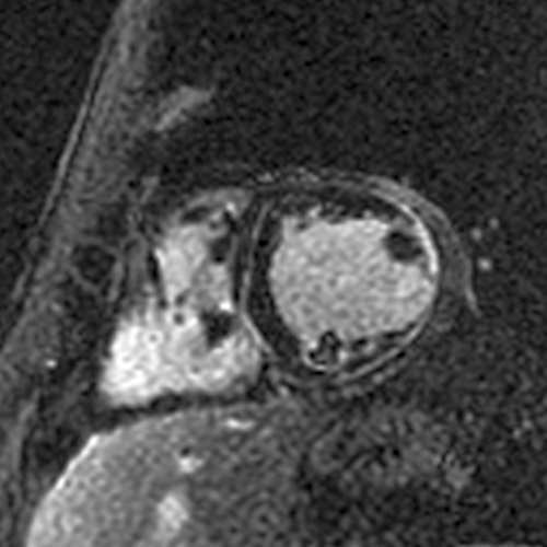
In the septum, the LGE spares the very RV side of the septum (also a distinguishing feature from cardiac sarcoidosis) and is therefore often midmyocardial… the ring is subepicardial laterally and midmyocardial septally.
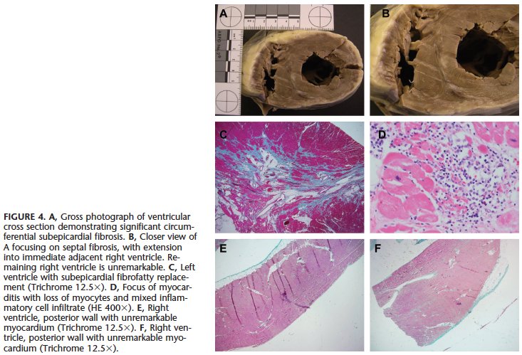
This pattern has been shown in ACM in pathology studies such as these:
ahajournals.org/doi/10.1161/CI…
pubmed.ncbi.nlm.nih.gov/19465822/
And CMR studies such as this:
academic.oup.com/ehjcimaging/ar…
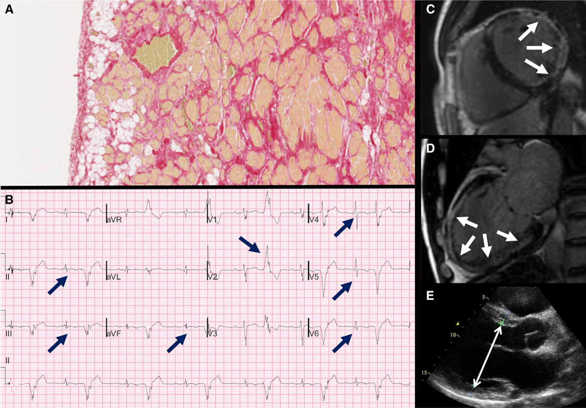
Elevated T1/T2 indicates an acute process (myocarditis), but it does not indicate the *cause*. We know ACM can present with acute myocarditis... this is one recent study:
ahajournals.org/doi/full/10.11…
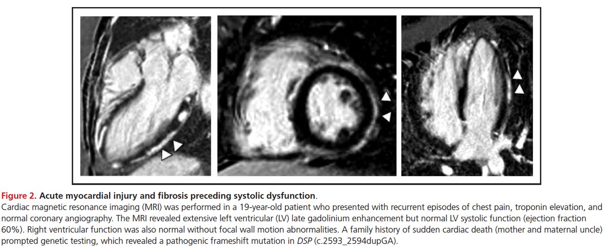
The fact that genetic ACM can present with myocarditis is not new... it has been described for 15-20 years. For example, here’s a sentence from a 2008 paper from the legendary Bill McKenna:
sciencedirect.com/science/articl…
Resolution of LGE does not tell us the cause of the myocarditis… it is simply a function of the extent of acute injury. Acute injury of a smaller extent can heal and become undetectable by LGE (= the scar is under the threshold of detection).
Bottomline – a diagnosis of myocarditis (like a diagnosis of NICM) is not wrong, but it’s incomplete. Not all myocarditis is viral.

We have a growing database of ACM cases, some with myocarditis... we hope to publish our experience at some point.

• • •

Missing some Tweet in this thread? You can try to force a refresh
 

Keep Current with Chetan Shenoy

Chetan Shenoy Profile picture

Stay in touch and get notified when new unrolls are available from this author!

Read all threads

This Thread may be Removed Anytime!

PDF

Twitter may remove this content at anytime! Save it as PDF for later use!

Try unrolling a thread yourself!

how to unroll video
  1. Follow @ThreadReaderApp to mention us!

  2. From a Twitter thread mention us with a keyword "unroll"
@threadreaderapp unroll

Practice here first or read more on our help page!

More from @cshenoy3

29 Apr
I previously did a thread on what the disappearance of LGE means. I’ll share two cases to illustrate some of my thoughts…

The first is a case of Desmoplakin cardiomyopathy that we published a few years ago:
ahajournals.org/doi/10.1161/CI…
A CMR 4 months later showed a near resolution of the anteroseptal/anterior/anterolateral LGE (red arrows). The inferior LGE (blue arrow) that did not disappear matched the fat seen on cine images. Image
Read 11 tweets
19 May 20
It’s been over 2 months since the first descriptions of cardiac manifestations of Covid-19. There have been many papers and reviews on this topic. What have we learned about how SARS-CoV-2 can affect the heart? #whyCMR #cardiotwitter
Troponin elevations and low EFs are frequently described. Why do they happen? Most papers use the term Covid-19 myocarditis. But can SARS-CoV-2 cause fulminant myocarditis (= extensive focal myocardial necrosis, as seen with viral lymphocytic or giant cell myocarditis)?
I tried looking at published CMR images and autopsy studies to get some insights into what happens in the hearts of Covid-19 patients. I’ll share my thoughts on 10 papers with CMR images and 2 papers each with autopsy data in >10 patients:
Read 28 tweets
12 Nov 19
Here’s an interesting paper published in JACC yesterday. The investigators studied 187 acute myocarditis patients with CMR (within a week) and repeated the CMR at 6 months.
onlinejacc.org/content/74/20/…
They found that LGE was present in 96% at the initial presentation and 86% at 6 months. They conclude:

“In the acute setting, LGE does not mean definite fibrosis, and it may disappear at 6 months.”
In the main text, they elaborate:

“Our data demonstrated that LGE in the acute phase of myocarditis is not necessarily synonymous with irreversible damage, because in 11% of our patients, LGE completely disappeared at follow-up.”
Read 13 tweets
24 Aug 19
With all the discussion about viability in the past few days, I would like to share how I interpret and report viability on CMR. I first look for LGE. Rarely, there’s no LGE and it’s all viable or more likely, a non-ischemic cardiomyopathy. #WhyCMR 1/18
When I see LGE, I confirm it’s in an ischemic pattern – subendocardial or transmural, and limited to a coronary territory, i.e., an MI. If not, it's again a non-ischemic cardiomyopathy and not a viability issue anymore. 2/18
Next, I try to identify how many and where the MIs are. For this, I look at the extent and locations of ischemic LGE and decide which of the 17 LV segments are likely to be supplied by each of the coronary arteries. 3/18
Read 18 tweets
15 Jul 18
@jameschilee @purviparwani @krychtiukmd @DrRyanPDaly @venkmurthy @journalofCMR @MarcDweck @AmitRPatelMD @onco_cardiology @ash71us @SHummelMD @SCMRorg 1/11 Why CMR for newly diagnosed HF? First, CMR is the gold standard for LVEF assessment and LVEF is used to diagnose cardiomyopathy, to decide medications and other treatments such as ICD and CRT. Sometimes, we don’t find a cardiomyopathy on CMR that was diagnosed on echo.
@jameschilee @purviparwani @krychtiukmd @DrRyanPDaly @venkmurthy @journalofCMR @MarcDweck @AmitRPatelMD @onco_cardiology @ash71us @SHummelMD @SCMRorg 2/11 Second, CMR tells us about the etiology of the cardiomyopathy. This is critical because the etiology is closely linked to prognosis and management. The prognostic relevance was nicely shown by @dukehfdoc in a landmark NEJM paper 18 years ago - goo.gl/xQP6JT
@jameschilee @purviparwani @krychtiukmd @DrRyanPDaly @venkmurthy @journalofCMR @MarcDweck @AmitRPatelMD @onco_cardiology @ash71us @SHummelMD @SCMRorg @DukeHFDoc 3/11 A 2016 AHA Scientific Statement also stresses the importance of the etiology of cardiomyopathy - goo.gl/cyoH2E
Treatments can be uniquely different for the various types of NICM – immunosuppression for sarcoidosis, eosinophilic and giant cell myocarditis...
Read 11 tweets

Did Thread Reader help you today?

Support us! We are indie developers!


This site is made by just two indie developers on a laptop doing marketing, support and development! Read more about the story.

Become a Premium Member ($3/month or $30/year) and get exclusive features!

Become Premium

Too expensive? Make a small donation by buying us coffee ($5) or help with server cost ($10)

Donate via Paypal Become our Patreon

Thank you for your support!

Follow Us on Twitter!