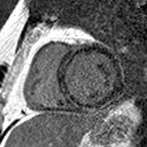
A CMR 4 months later showed a near resolution of the anteroseptal/anterior/anterolateral LGE (red arrows). The inferior LGE (blue arrow) that did not disappear matched the fat seen on cine images.
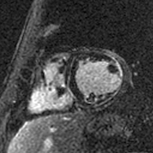
All short axis LGE images at the time of the acute myocarditis.
All short axis LGE images 4 months later showing a significant decrease in the LGE.
Next case - this patient had a STEMI with chest pain, anterior ST elevations, and elevated troponin. Coronary angiography showed disease in the LAD that was felt to be the culprit and was treated with 3 stents.
CMR during the hospitalization for the STEMI showed a patchy MI in the LAD territory. CMR 6 months later showed no LGE. Small/patchy MIs can also disappear.
All short axis LGE images at the time of the STEMI.
All short axis LGE images 6 months later showing the disappearance of the LGE.
This JACC Scientific Expert Panel article led by @Borjaibanez1 includes a nice cartoon explaining why patchy MIs can “disappear”.
Bottomline – The disappearance of LGE does not tell us the cause of the cardiomyopathy… it simply means a small amount of heart muscle died from the acute process (myocarditis due to any cause, MI, etc.)
I think it’s genetic ACM because of the pattern of LGE involvement. There is circumferential LGE (the "ring sign") with lateral wall predominance vs. septal (unlike in cardiac sarcoidosis).
The lateral wall involvement spares the most subendocardial portion and/or the papillary muscles and trabeculations, indicating it is subepicardial (unlike a transmural MI that started subendocardially and involves the papillary muscles/trabeculations).
In the septum, the LGE spares the very RV side of the septum (also a distinguishing feature from cardiac sarcoidosis) and is therefore often midmyocardial… the ring is subepicardial laterally and midmyocardial septally.
It’s been over 2 months since the first descriptions of cardiac manifestations of Covid-19. There have been many papers and reviews on this topic. What have we learned about how SARS-CoV-2 can affect the heart? #whyCMR#cardiotwitter
Troponin elevations and low EFs are frequently described. Why do they happen? Most papers use the term Covid-19 myocarditis. But can SARS-CoV-2 cause fulminant myocarditis (= extensive focal myocardial necrosis, as seen with viral lymphocytic or giant cell myocarditis)?
I tried looking at published CMR images and autopsy studies to get some insights into what happens in the hearts of Covid-19 patients. I’ll share my thoughts on 10 papers with CMR images and 2 papers each with autopsy data in >10 patients:
Here’s an interesting paper published in JACC yesterday. The investigators studied 187 acute myocarditis patients with CMR (within a week) and repeated the CMR at 6 months. onlinejacc.org/content/74/20/…
They found that LGE was present in 96% at the initial presentation and 86% at 6 months. They conclude:
“In the acute setting, LGE does not mean definite fibrosis, and it may disappear at 6 months.”
In the main text, they elaborate:
“Our data demonstrated that LGE in the acute phase of myocarditis is not necessarily synonymous with irreversible damage, because in 11% of our patients, LGE completely disappeared at follow-up.”
With all the discussion about viability in the past few days, I would like to share how I interpret and report viability on CMR. I first look for LGE. Rarely, there’s no LGE and it’s all viable or more likely, a non-ischemic cardiomyopathy. #WhyCMR 1/18
When I see LGE, I confirm it’s in an ischemic pattern – subendocardial or transmural, and limited to a coronary territory, i.e., an MI. If not, it's again a non-ischemic cardiomyopathy and not a viability issue anymore. 2/18
Next, I try to identify how many and where the MIs are. For this, I look at the extent and locations of ischemic LGE and decide which of the 17 LV segments are likely to be supplied by each of the coronary arteries. 3/18