#FOAMrad #neurorad #radres #radmed #HUNSC #daybydaycases #radiology #MedEd
One patient/two sides……and two pathologies.
From left to right axial FLAIR, coronal T2 and axial T1 MRI
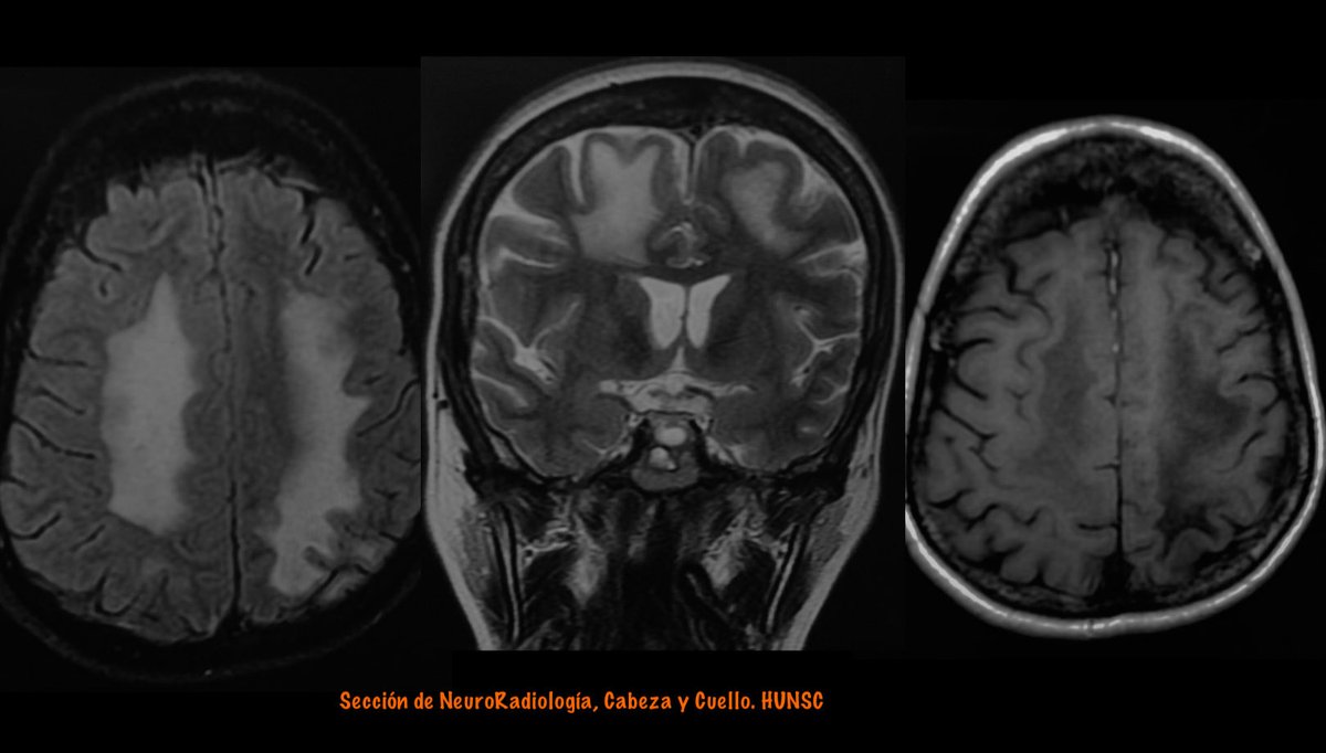
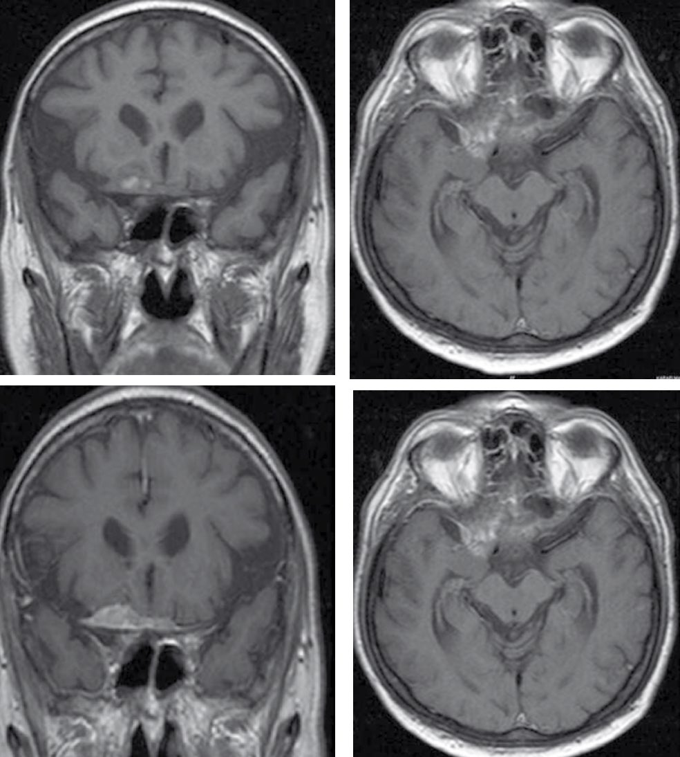
Right side:
Parasagital meningioma with vasogenic edema
From left to right: coronal T2, enhanced T1 and DWI.
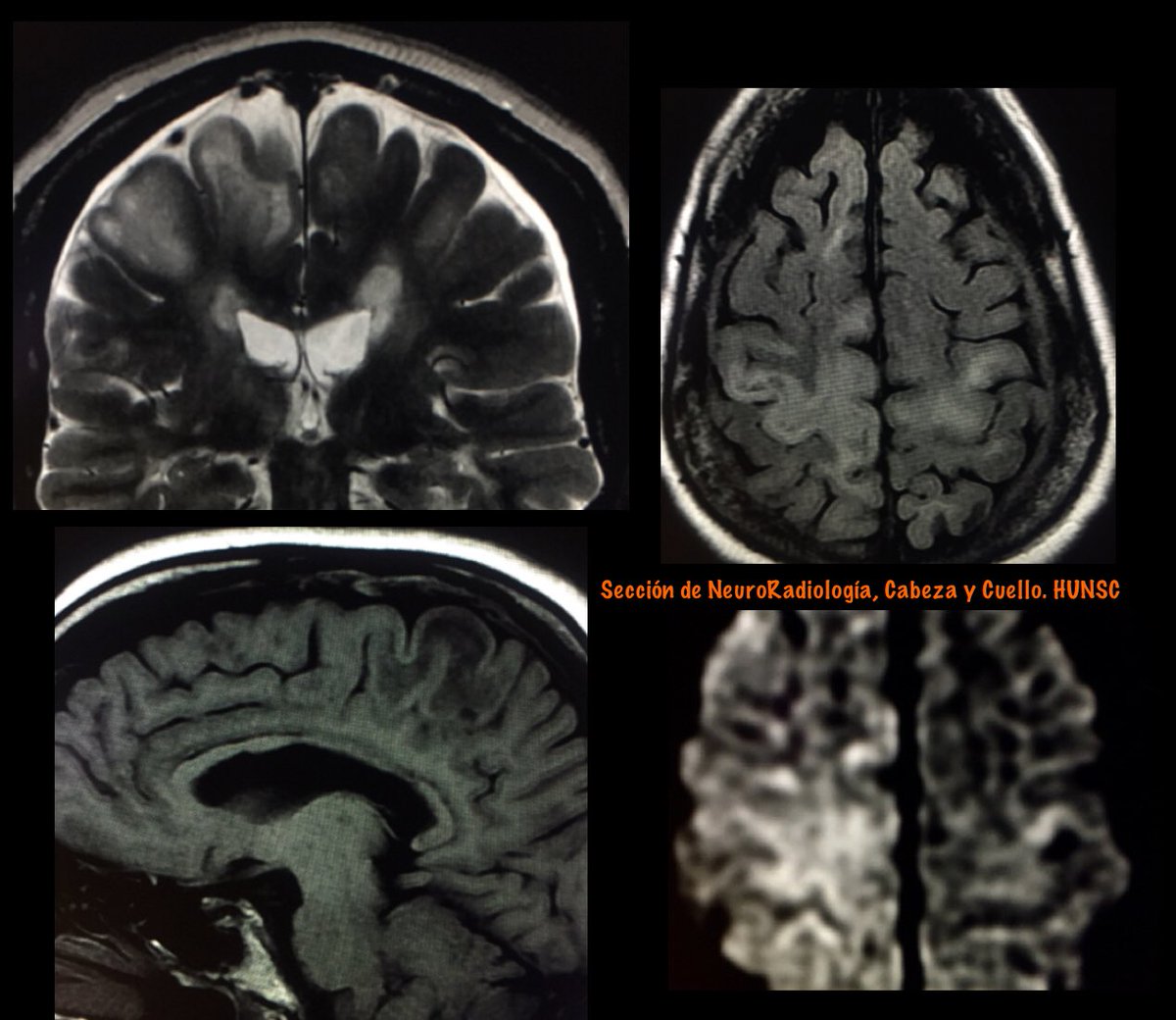
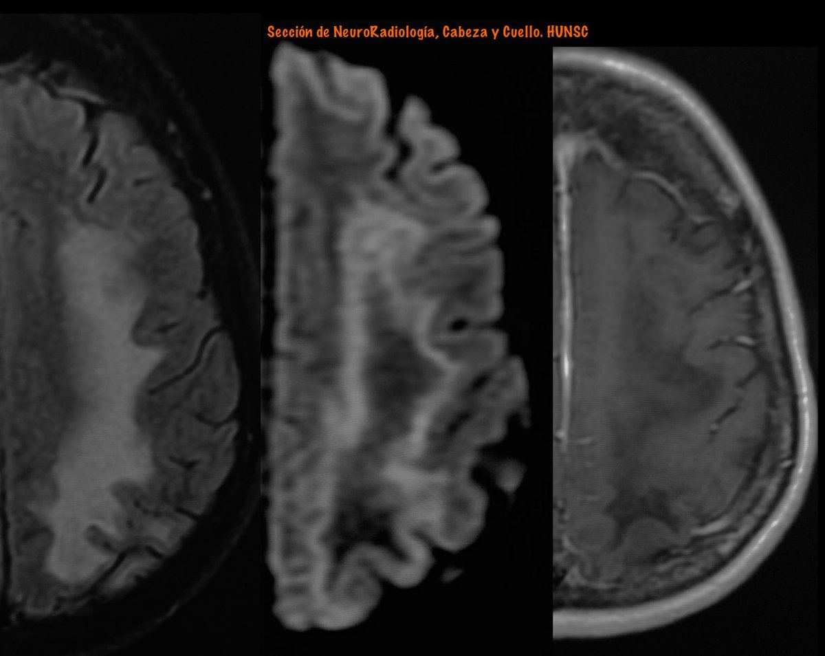
Left side :
Progressive Multifocal Leukoencephalopathy (PML)
From Left: FLAIR, DWI and enhanced T1 MRI
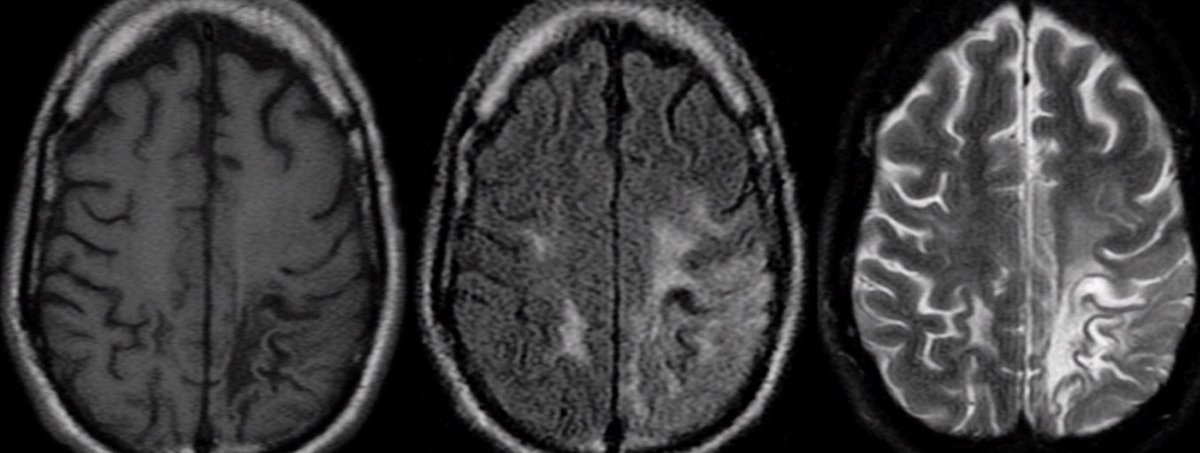
Diffusion Imaging In PML:
The appearance on DWI varies according to the disease stage.
In new active lesions, there is a rim of diffusion restriction at the advancing edge and a central core of facIlitated diffusion.
The rim is usually incomplete and signifies active infection.
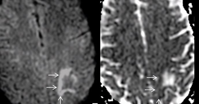
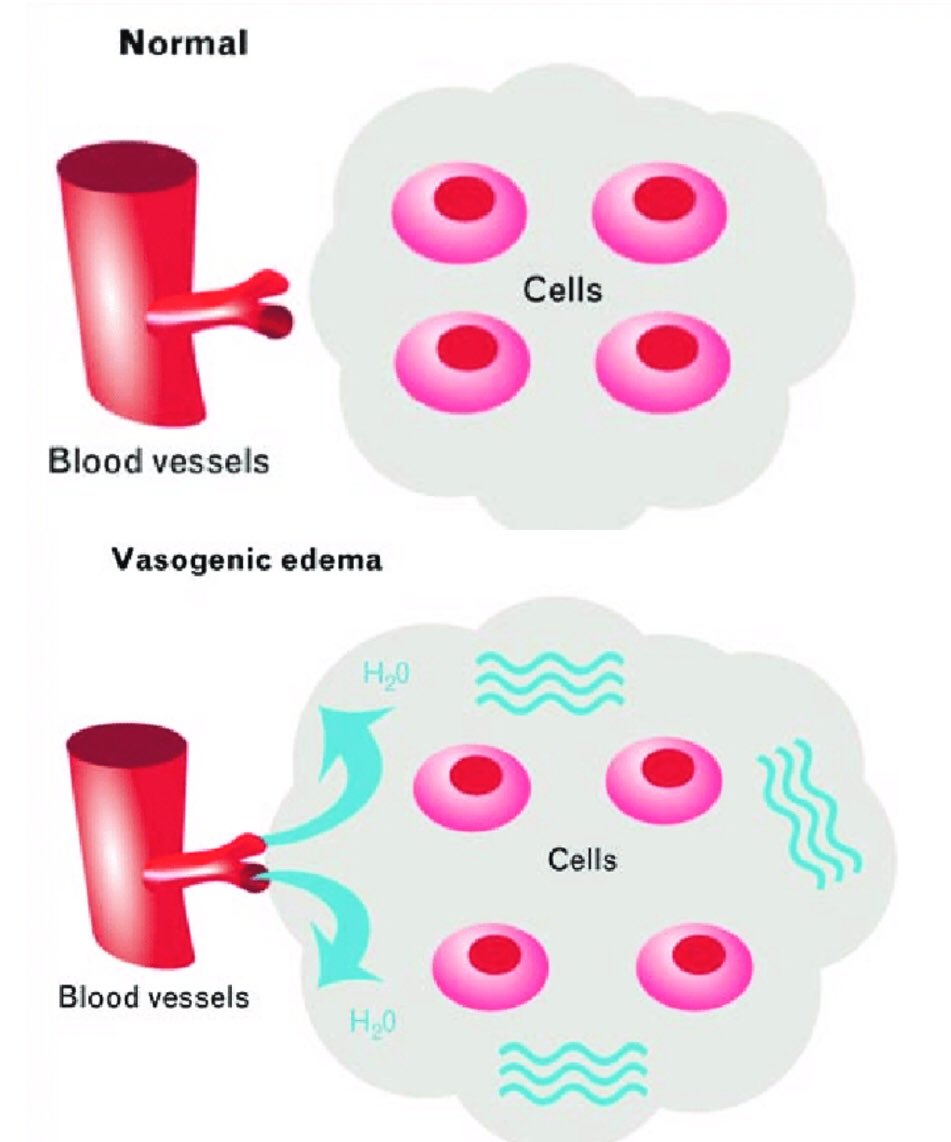
Histopathologically, this advancing edge correlates with large swollen oligodendrocytes, enlarged “bizarre astrocytes” with numerous large processes, and infiltration of foamy macrophages. This cellular enlargement constricts the extracellular space.
AJNR 2010
DOI 10.3174/ajnr.A2035

10.1594/ecr2015/B-0300

Neurology 2016;86;1516-1523
DOI 10.1212/WNL.0000000000002586

AJNR 2015
dx.doi.org/10.3174/ajnr.A

Curr Opin Neurol 2014, 27:260–270
DOI:10.1097/WCO.0000000000000099

• • •

Missing some Tweet in this thread? You can try to force a refresh
 

Keep Current with Vicente Martín

Vicente Martín Profile picture

Stay in touch and get notified when new unrolls are available from this author!

Read all threads

This Thread may be Removed Anytime!

PDF

Twitter may remove this content at anytime! Save it as PDF for later use!

Try unrolling a thread yourself!

how to unroll video
  1. Follow @ThreadReaderApp to mention us!

  2. From a Twitter thread mention us with a keyword "unroll"
@threadreaderapp unroll

Practice here first or read more on our help page!

More from @vmargar

3 Feb 20
#FOAMrad #neurorad #HUNSC #daybydaycases
Two patients with a similar clinical profile: elderly men with prostatic adenocarcinoma.
Similar neuroimaging pattern: dural base lesion with intense enhancement and vasogenic edema.
But with different etiology .....
A: Meningioma
B: Dural metastases (DM)
DM in isolation is rare, comprising <1% of all intracranial metastases, and most commonly originates from renal, lung, and breast cancer, as well as carcinoid, adenoid cystic carcinoma, prostatic adenocarcinoma (PCa), and dermatofibrosarcoma
Transdural metastases may mimick meningiomas in radiologic imaging, especially in patients with undiagnosed symptoms of prostatism or PCa.
In older men with dural lesion(s), a possibility of PCa metastases must be considered and radiologic evaluation
Read 4 tweets
12 Aug 19
#FOAMrad #radres #neurorad
Cerebral Edema
Is an excess accumulation of water in the intracellular and/or extracellular spaces of the brain
Is a common response to various forms of brain injury, and the causes can be categorized as cytotoxic, vasogenic, interstitial, or combined
Cytotoxic edema
• Is cell swelling caused by damaged molecular sodium–potassium ATPase ion pumps. It can affect both gray and white matter.
• Is caused by cell death, most commonly due to infarct or excitotoxic (secondary to excessive neurotransmitter stimulation) brain injury.
Vasogenic edema
• Is interstitial edema caused by increased capillary permeability, by breakdown of the tight endothelial junctions comprising the BBB, secondary to either physical disruption or release of vasoactive compounds
• It is seen primarily in the white matter
Read 5 tweets
14 Jun 19
PML in MS
Progressive multifocal leukoencephalopathy (PML) is an opportunistic infection of the CNS caused by John Cunningham virus (JCV)
PML in the setting of monoclonal antibody therapy has attracted tremendous attention particularly in MS patients treated with natalizumab/NTZ. Image
Features favoring PML over MS (as proposed by Yousry et al) are the following:
1) Diffuse subcortical rather than periventricular white matter involvement; frequent involvement of posterior fossa.
2) Irregular ill-defined infiltrating edge confined to the white matter. Image
3) Persistent progression of the lesion confined within the white matter tract.
4) No mass effect even in large lesions.
5) Diffuse increased T2 signal intensity; recently involved areas more T2 hyperintense than the old areas. Image
Read 8 tweets

Did Thread Reader help you today?

Support us! We are indie developers!


This site is made by just two indie developers on a laptop doing marketing, support and development! Read more about the story.

Become a Premium Member ($3/month or $30/year) and get exclusive features!

Become Premium

Too expensive? Make a small donation by buying us coffee ($5) or help with server cost ($10)

Donate via Paypal Become our Patreon

Thank you for your support!

Follow Us on Twitter!