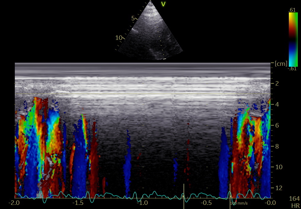
2/ 22 healthy subjects, Valve openings and closures timed by Doppler flow, and transewfrred to Tissue Doppler recordings.
3/ Pre ejection velocities started 24.8 ms after start QRS, with a duration of 51.5 ms, ending about 11.5 ms after MVC. Thus, both electromechanical delay and pre ejection velocity occurs *before* onset of IVC, and are not a measure of IVC or Isovolumic acceleration.
4/ This has been shown by ultra high frame rate imaging previously. So what is the significance of the pre ejection velocities? It's not recoil from atrial relaxation, as it's present in fib, and absent in AV-block. pubmed.ncbi.nlm.nih.gov/24210859/
5/ Both initial pressure increase pubmed.ncbi.nlm.nih.gov/623315/ and volume decrease pubmed.ncbi.nlm.nih.gov/18606917/ occurs before MVC, indicating that the pre ejection velocities are active contraction. The latter reference has shown it also by stenting the MV.
6/ In the present study, we show the active shortening by strain rate. Thus, this seems to be the marker of electromechanic activation, which is simultaneous in septum and the lateral wall, possibly by left anterior and left posterior bundle.
7/ This has been shown previously: electrical breakthrough simultaneous in septum and laterally pubmed.ncbi.nlm.nih.gov/6723010/ , and with an electromechanical delay of about 20 - 30 ms pubmed.ncbi.nlm.nih.gov/14670817/ , in line with our study: 25 ms from onset Q to onset pre ejection spike
8/ But what is then the significance of the pre ejection contraction?
-firstly, a small volume reduction, as seen by mitral annulus displacement. Not due to ejection, but exclusion of the volume by the annulus.
9/ - Secondly, as the annulus moves in a stationary blood column, the leaflets will be pushed towards closure, possibly in combination by the last of the filling vortex, so pre ejection contraction is part of the mechanism for MVC.
10/ -Thirdly, the apical motion will stop at the closure of the MV, which is the start of the real IVC. Including the pre ejection spike in IVC will over estimate IVC by about 40 ms, and including EMD as well, will over estimate IVC by further 25ms.
11/ There is no ring motion during IVC, LV being isovolumic, and concepts like IVC velocity or acceleration are erroneous. Start ejection is at AVO, when LV and Ao pressures are equalised, the start of ring motion during ejection is, of course simultaneous in all parts
12/ -and the onset of the main shortening event, is the end of IVC/AVO, and certainly not electromechanical activation.
• • •
Missing some Tweet in this thread? You can try to
force a refresh
1/ Man ca 30, exertional dyspnea, CPET with normal VO2max, but pulmonologist concerned about possible drop in CO at peak exercise. Normal resting echo, no LVOT obstruction or gradient, no MR. Dobutamine stress: Chordal SAM, no regional ischemia
2/ Intraventricular gradient
3/ shown by CMM to be mid ventricular, moving towards apex in systole. No concomitant MR.
What next?
1/ #LBBB generates often a classical pattern on #EchoFirst. The pattern is very distinctive in Tissue Doppler of the septum.
The classical pattern arises from the time lapse of the activation and relaxation of the two walls, creating a pattern of interaction due to a sequence temporal imbalances of the tension between the two walls.
2/ As the septum is activated first, it contracts (shortening - septal flash) without activation of the lateral wall, which stretches. This generates slower pressure build up than a normal IVC, which then is prolonged.
It's the ratio of SV and BP that's closest to afterload dependence. MW being the *product* of SV (and hence also preload dependent) is very afterload dependent (although an inverted U relation can be hypothetisized.
3/ Thus, if SV ⬇️, MW⬇️, but if LVEDV also ⬇️, EF will ➡️. Sp far so good. But if BP ⬆️, MW⬆️, even if SV and GLS ⬇️ and EF➡️, as shown in pubmed.ncbi.nlm.nih.gov/32966690/, meaning that the demand increased while performance (SV) decreased, both due to increased afterload.
1/ Thread on myocardial work. What does it actually mean, and is it really useful? It is a spin-off from pressure-volume loops, which are an illustration to visualise the relation between stroke volume, pressure and contractility, and to assess physiology in animal experiments.
2/ The area of the PV-loop is LV ejection work. The height of the PV-loop is the SBP-LVDBP difference, the width is the SV. Mean SBP and mean LVDBP ((blue dotted rectangle), shows the relation in an easy way. GMW is SV x (mean SBP - mean LVDBP).
3/ This, of course means that it is definitely preload dependent, as increased preload increases SV. Increased afterload, on the other hand increases pressure work, but as afterload decreases SV, the relation is somewhat more uncertain.
Our last paper is out: Left ventricular longitudinal shortening: relation to stroke volume and ejection fraction in ageing, blood pressure, body size and gender in the HUNT3 study openheart.bmj.com/content/7/2/e0…
I'll discuss some of the findings and their implications in a thread
2/ 1266 subjects without history of HT, heart disease or diabetes, LV linear measurements of systolic and diast. wall thickness, length, and diameters, entered into an ellipsoid LV model.
3/ This gave us the possibility to look at age dependent changes in both volumes and functional measures, but with the two main limitations of the cross sectional nature of the study and for volumes the limitations of geometric model itself. There are still non-resolved issues.
1/ Tweetorial. Prompted by a question of "layer strain", I'd like to go into that, as the concept is based on a completely erroneous perception of strain components somehow related to the directional fibre shortening. This is not the case.
2/ The three normal strains are longitudinal, circumferential and transmural (or radial). The relations between all three major strains are explored in the HUNT study: openheart.bmj.com/content/openhr…
3/ Transmural (radial) strain is simply wall thickening, while circumferential strain is fractional circumferential shortening, which, as the circumference is 3.14*diameter, equals fractional diameter shortening.