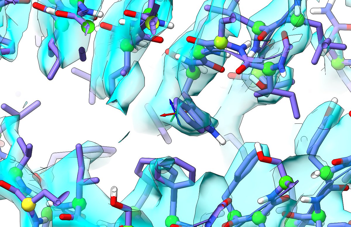
For your delectation: the Rhodobacter sphaeroides LH2 antenna complex at a glorious 2.1 Angstrom resolution. Building this one was an absolute joy, with maps (courtesy of new-to-Twitter @PuQian3) so good they almost felt like cheating! (1/7)
Remember the big RC-LH1 dimer complex from a few weeks back? These smaller LH2 complexes act as little satellites to that, increasing the light-gathering area and passing their excitation energy back to the LH1 complex. (2/7)
My favourite part of this model was figuring out this site at the N-terminus of the alpha chain. Yes, that *is* the N-terminus. It's what's known as N-carboxymethionine, and it's what you get when nature desperately wants an acid but only has an amine to work with. (3/7)
This has been seen in this context once before in a related bacterium, in a crystal structure published back in 2003 (1nkz) - but its presence in Rba. sphaeroides was uncertain. Similar carboxylation of lysine sidechains also happens from time to time... (4/7)
It's a spontaneous, non-enzymatically driven post-translational modification - basically, the amine captures a carbon dioxide molecule to form the carbamate because that's what the local environment really "wants". (5/7)
It's also a beautiful example of a modification you'll (probably) only ever discover by capturing the structure experimentally. Since it's entirely environment-driven there's no sequence motif to look for; it decomposes in acid making it invisible to HPLC/MS... (6/7)
... but once you see it in the density it's really quite unmistakable (and beautiful). Anyway, for those still with me here's a fly-through of all residues in the beta chain (wireframe at 6.6 sigma, surface at 10 sigma). (fin)
*alpha chain. Sigh... I’m off to bed.
• • •
Missing some Tweet in this thread? You can try to
force a refresh
There's (quite rightly) a lot of raving about the new AlphaFold DB resource today. Thought I'd weigh in with my own example. DNA-dependent protein kinase catalytic subunit (uniprot.org/uniprot/P78527) is an enormous beast - a single chain of 4,128 residues! (1/10)
There are now 19 structures of this guy in the wwPDB, all post-resolution-revolution cryoEM structures. The first (2017) was reconstructed to a character-building 4.3 Å. I'm told the model building was extremely challenging, ... (2/10)
... and a glance at the overall structure shows why. The thing is almost entirely composed of short helix after short helix after short helix... without clear sidechain information getting out of step with the map would be really, really easy. (3/10)
As we all know by now, the most pressing current concern about the ongoing pandemic is the potential for emergence of immune-escape variants. With that in mind, I'm very happy to have had the chance to contribute to this important work: (1/6) nature.com/articles/s4158…
A major multi-centre effort including @tylernstarr, @jbloom_lab, @Vir_Biotech, @jchodera, @ivyzhang__, @W_Glass and many others, this work assesses a wide panel of antibodies against the SARS-CoV-2 spike for potency, breadth and potential for immune escape. (2/6)
While the potency of many existing antibodies is dramatically reduced by mutations tolerated by the virus itself, the great news is that a few show high potency against most or all known sarbecoviruses. (3/6)
A more extreme illustration of the problem: yes, this is a single residue (JSG, found in 6s8h and 6mhu). What it actually *is* is E. coli lipopolysaccharide (LPS) - how it appears in the database is {deep breath}:
As is becoming increasingly common these days, the pace of reality has well outstripped that of the scientific publication cycle. Still, I'd like to share this with you: sciencedirect.com/science/articl… (1/14)
Unless you've been entirely disconnected for the last month or three, the core message (that immunity-escaping variants of SARS-CoV-2 are developing and need to be watched) should come as no surprise. This work focuses on one of the first such mutants identified, N439K. (2/14)
At the time of submission, the N439K variant had been spotted in 34 different countries and had arisen independently multiple times, suggesting at least some gain of fitness over the wild-type virus. (3/14)
ISOLDE 1.0 is finally live! To get it, just install ChimeraX 1.0 from rbvi.ucsf.edu/chimerax/downl…, then go to Tools/More Tools..., find ISOLDE and click Install. In the thread, I'll give a quick recap of what ISOLDE is, followed by a rundown of what's new. (1/17)
So what is ISOLDE? In brief, it's an interactive environment for (re)building atomic models into medium-low-density crystallographic and cryo-EM maps using GPU-accelerated interactive molecular dynamics. That's a bit of a mouthful, so here's a video demo in the next tweet. (2/17)
This example (found this morning) shows the correction of an out-by-one error in a beta strand (residues 306-318 of 3mca chain B). Like all videos in this thread, it's an actual-speed screen capture. Key features to note: (3/17)
In all cryo-EM maps of the SARS-CoV-2 spike protein density for the N-terminal domain has been really rubbishy, with modelling only really possible based on somewhat-weak homology to the original SARS equivalent... until now. (1/12)
On the left: a better-resolved region from this domain in 6vxx - general path of the backbone is fine, but sidechains are uninterpretable without outside info. The outer surface loops devolve to complete rubbish. On the right, the same site from 6zge. Clear, unambiguous (2/12)
So what caused this enormous difference in quality? I asked the authors (from the lab of Steve Gamblin at the Crick), and their honest answer was that they weren't sure - but they pointed me at this intriguing preprint from Christiane Schaffitzel: biorxiv.org/content/10.110… (3/12)