
#IBDtwitter
In ileocecal #Crohn’s disease, typically all bowel wall layers are involved. #IUS shows marked mural thickening, predominantly in the terminal ileum, cecum & appendix can also be involved.
🔗: ncbi.nlm.nih.gov/pmc/articles/P…
In ileocecal #Crohn’s disease, typically all bowel wall layers are involved. #IUS shows marked mural thickening, predominantly in the terminal ileum, cecum & appendix can also be involved.
🔗: ncbi.nlm.nih.gov/pmc/articles/P…

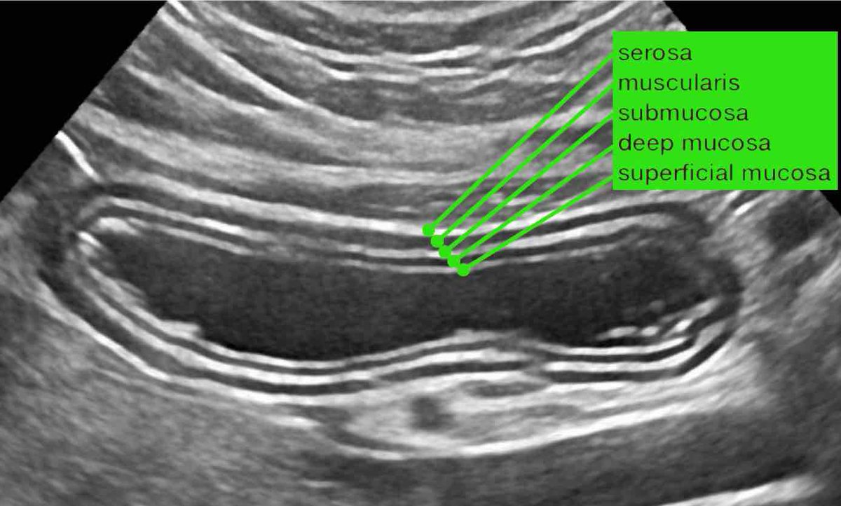
Bowel wall thickening is most prominent in the hyperechoic submucosal layer, representing deposition of fat and fibrous tissue as a result of chronic bowel inflammation

Difference between size and compressibility of normal ileum (left) and #Crohn affected ileum during intermittent graded compression. #IBDtwitter


Using graded compression, two adjacent bowel segments are compressed against the iliopsoas muscle.
The ventral segment is normal and is easily compressed, while the Crohn’s loop can hardly be compressed. #IBDtwitter #GITwitter
The ventral segment is normal and is easily compressed, while the Crohn’s loop can hardly be compressed. #IBDtwitter #GITwitter


Measuring bowel wall thickness is best & most reproducibly performed during compression, normal (left) & Crohn’s (right) Measurements are from outer contour of the muscular layer to the opposite side & then divided by 2 resulting in wall thickness of 1.5 mm & 6.5 mm for normal

At first presentation, #IUS already reveals the characteristic transmural “signature” of Crohn’s disease. This transmural progression is recognized as focal, hypoechoic changes (*) in the otherwise hyperechoic submucosa, closely correlating with endoscopic findings

Case: This 18-yr old man was admitted with acute appendicitis-like symptoms & immediate CT was done. CT revealed evident ileal bowel wall thickening and a normal appendix. Subsequent #IUS showed transmural hypoechoic changes in the hyperechoic submucosa confirming Crohn’s ileitis

Patients with active #Crohn’s disease, the normal #IUS bowel wall may be lost diffusely.
👀 hyperechoic fatty tissue (fat) around the ileum, representing the inflamed mesentery & omentum, trying to wall off the imminent perforation. #IBDtwitter #GITwitter
👀 hyperechoic fatty tissue (fat) around the ileum, representing the inflamed mesentery & omentum, trying to wall off the imminent perforation. #IBDtwitter #GITwitter

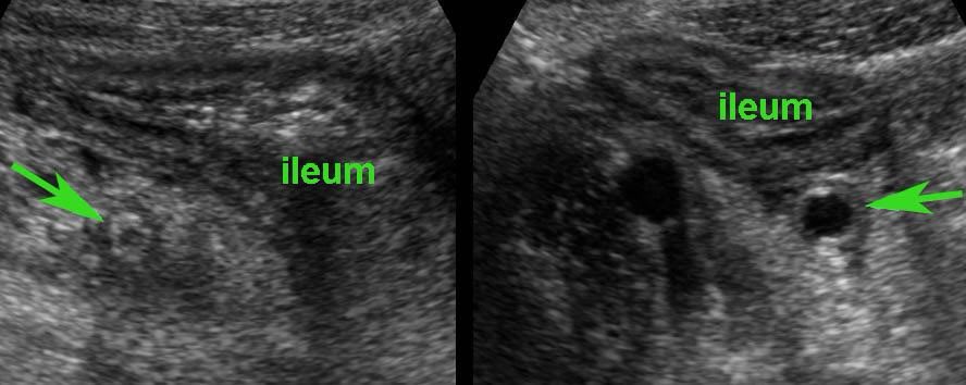
One features of Crohn’s disease is the patchy way it affects bowel.
This results in #skiplesions, where large parts of the bowel are left unharmed
The affected parts show a relatively sharp demarcation, the transition from normal (small arrows) to abnormal bowel (large arrows)
This results in #skiplesions, where large parts of the bowel are left unharmed
The affected parts show a relatively sharp demarcation, the transition from normal (small arrows) to abnormal bowel (large arrows)

Bright #eccentric foci within the hypoechoic areas are air-configurations representing deep #ulceration, here depicted in two different patients.
These #transmural ulcerations herald sinus tract formation, abscesses & #fistula formation
#IBDtwitter #GITwitter
These #transmural ulcerations herald sinus tract formation, abscesses & #fistula formation
#IBDtwitter #GITwitter

#IBDtwitter #GITwitter
Air-configurations reaching beyond the peripheral borders of the bowel wall definitely indicate sinus tract formation in 2 different patients
Air-configurations reaching beyond the peripheral borders of the bowel wall definitely indicate sinus tract formation in 2 different patients

#Abscess
Actively inflamed terminal ileum with a moderately demarcated hypoechoic area outside the bowel wall, surrounded by inflamed fat, indicates sinus tract and abscess formation #IBDtwitter #GITwitter
Actively inflamed terminal ileum with a moderately demarcated hypoechoic area outside the bowel wall, surrounded by inflamed fat, indicates sinus tract and abscess formation #IBDtwitter #GITwitter

2 patients with Crohn’s abscesses close to the ileum.
Abscesses in Crohn’s disease are often small and collapsed.
Abscesses usually have an open connection to the ileal lumen, allowing pus to immediately evacuate to the bowel lumen when pressure goes up #IBDtwitter #GITwitter
Abscesses in Crohn’s disease are often small and collapsed.
Abscesses usually have an open connection to the ileal lumen, allowing pus to immediately evacuate to the bowel lumen when pressure goes up #IBDtwitter #GITwitter

Ultimately sinus tract formation may proceed to fistula formation.
Here 2 examples of entero-enteric fistulization (arrowheads) #IBDtwitter #GITwitter
Here 2 examples of entero-enteric fistulization (arrowheads) #IBDtwitter #GITwitter

Fistula (arrowheads) from the terminal ileum to the appendix (arrows) in advanced ileocecal Crohn’s disease.
Hyperechoic fat, surrounding the fistula complex, representing omentum and mesentery effectively walling off a possible imminent perforation to the peritoneal cavity
Hyperechoic fat, surrounding the fistula complex, representing omentum and mesentery effectively walling off a possible imminent perforation to the peritoneal cavity

Ultimately sinus tract and abscess formation may result in a fistula.
The most frequent targets are small bowel, sigmoid and appendix.
Unusually, fistula to the bladder, the abdominal wall and iliopsoas muscle may occur.
The most frequent targets are small bowel, sigmoid and appendix.
Unusually, fistula to the bladder, the abdominal wall and iliopsoas muscle may occur.


In situations with complex enterocutaneous fistula involving multiple small bowel loops, CT or MRI enterography is the ideal modality for pre-operative mapping and in follow-up.
Stenosis and prestenotic dilatation
Inflammatory bowel wall thickening or the ensuing fibrotic strictures may finally result in luminal narrowing, stenosis and bowel obstruction.
The affected segment typically demonstrates dysfunctional motility.
Inflammatory bowel wall thickening or the ensuing fibrotic strictures may finally result in luminal narrowing, stenosis and bowel obstruction.
The affected segment typically demonstrates dysfunctional motility.

#fibrotic stenosis in an ileal segment.
Note the prestenotic dilatation and absence of #peristalsis at the level of the #stricture (arrows).
Note the prestenotic dilatation and absence of #peristalsis at the level of the #stricture (arrows).

In stenosing #Crohn’s disease with long episodes of chronic recurrent #smallbowelobstruction, an interesting phenomenon in ileal loops may be observed & hypertrophy of the muscular layer.
This so-called “inflammation-smooth muscle hyperplasia axis”
This so-called “inflammation-smooth muscle hyperplasia axis”

In longstanding Crohn’s disease, “creeping fat” can be found, which is hypertrophied mesenteric fat “wrapped around” the affected bowel, most frequently in the ileocecal region.


Old and inactive Crohn’s disease of the terminal ileum in a now asymptomatic patient.
Note prominent thickening of the hyperechoic third layer, representing fatty deposition and loose connective tissue in the submucosa. #IBDtwitter #GITwitter
Note prominent thickening of the hyperechoic third layer, representing fatty deposition and loose connective tissue in the submucosa. #IBDtwitter #GITwitter

an example of appendiceal involvement in ileocecal Crohn’s disease.
Note prominent echolucent wall thickening of both terminal ileum and appendix (arrowheads). #IBDtwitter #GITwitter
Note prominent echolucent wall thickening of both terminal ileum and appendix (arrowheads). #IBDtwitter #GITwitter

#IUS reveals mucosal and submucosal thickening and hyperemia of the entire colon, which is impossible to differentiate from ulcerative colitis. Since in every patient with suspected IBD, repeated stool cultures are performed, the correct diagnosis is not a problem in most cases

As clinically observed in Campylobacter patients, pain in Campylobacter ileocecitis is often intermittent and crampy in nature.
During these crampy pain attacks (remember “Crampylobacter”), the ileum is slightly intussuscepted into the cecum, sliding back again after the attack
During these crampy pain attacks (remember “Crampylobacter”), the ileum is slightly intussuscepted into the cecum, sliding back again after the attack

Differentiating Crohn’s disease and infectious ileocolitis is usually easy, as in the latter the layer structure is virtually always intact and there is never inflamed fat, abscess or fistula formation

Tuberculous ileitis can closely mimic Crohn’s disease, and even show abscesses, fistula formation and fibrotic stenosis

Clostridium colitis may mimic the US features of ulcerative colitis, although the (sub)mucosal wall thickening in Clostridium colitis is usually much more prominent.
Clinical findings may at times be quite confusing
Clinical findings may at times be quite confusing

Ischemic colitis has a predilection for the splenic flexure of the colon.
#IUS findings of sharply demarcated, segmental, hypoechoic wall thickening with transmural features and fat stranding may closely mimic Crohn’s colitis, both on US and CT.
#IUS findings of sharply demarcated, segmental, hypoechoic wall thickening with transmural features and fat stranding may closely mimic Crohn’s colitis, both on US and CT.

Note the abrupt transition of normal and abnormal colonic wall (arrowheads) mimicking skip lesions as in Crohn’s disease.
Clinical findings as well as the atherosclerotic origin of the SMA (arrow) give the clue here.
Clinical findings as well as the atherosclerotic origin of the SMA (arrow) give the clue here.

Patients with segmental bowel wall thickening & loss of the normal #IUS, active Crohn’s disease may mimic malignancy. Not only the relatively hypoechoic tumor itself is not compressible, but also the surrounding hyperechoic fat (arrowheads), representing desmoplastic reaction

Irregular, circumferential wall thickening of the cecum with loss of layer structure & ill-defined borders, separating it from the surrounding inflamed fat.
CT scan, malignancy was considered more likely.
#Colonoscopy revealed cecal carcinoma.
CT scan, malignancy was considered more likely.
#Colonoscopy revealed cecal carcinoma.

Sometimes, the US image can be quite confusing.
The irregular, asymmetrical wall thickening of the cecum with adjacent inflamed fat (*) and encroachment of the appendix (arrow) suggested malignancy with desmoplastic reaction and ingrowth in the base of the appendix
The irregular, asymmetrical wall thickening of the cecum with adjacent inflamed fat (*) and encroachment of the appendix (arrow) suggested malignancy with desmoplastic reaction and ingrowth in the base of the appendix

An important pitfall is secondary thickening of the terminal ileum due to advanced appendicitis.
When the appendix is not visualized in such cases, secondary wall thickening of the ileum may be misinterpreted and may lead to serious delay of surgery.
In doubt, CT very helpful
When the appendix is not visualized in such cases, secondary wall thickening of the ileum may be misinterpreted and may lead to serious delay of surgery.
In doubt, CT very helpful
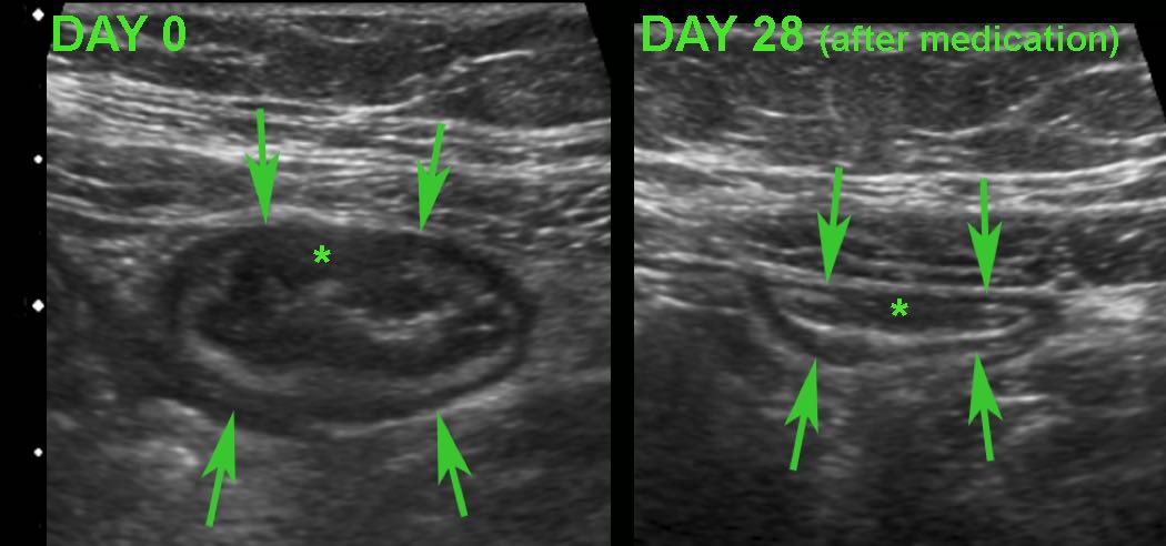
Monitoring disease activity in response to medical therapy at the level of the terminal ileum.
There is convincing decrease in wall thickening after 4 weeks of medication.
Note subtle residual hypoechoic, transmural changes (*) still visible in the near wall after therapy.
There is convincing decrease in wall thickening after 4 weeks of medication.
Note subtle residual hypoechoic, transmural changes (*) still visible in the near wall after therapy.
Demonstration of hypoechoic, transmural inflammation and the presence of non-compressible inflamed fat strongly favor Crohn’s disease #GITwitter #IBDtwitter

Severe ulcerative colitis ⬆️echogenicity of the surrounding fat (*)
This fat is rather well-compressible & should be considered as edema associated with secondary hyperemia rather than as a sign of true transmural inflammation
Wall thickening of the transverse colon (arrowheads)
This fat is rather well-compressible & should be considered as edema associated with secondary hyperemia rather than as a sign of true transmural inflammation
Wall thickening of the transverse colon (arrowheads)

#IBDtwitter #GITwitter
“Bowel US, a non-invasive and patient friendly procedure, may replace CS whenever mucosal biopsies are not required”
onlinelibrary.wiley.com/doi/10.1002/ue…
“Bowel US, a non-invasive and patient friendly procedure, may replace CS whenever mucosal biopsies are not required”
onlinelibrary.wiley.com/doi/10.1002/ue…
@DrMikeDolinger
“The use of the MUC at baseline to determine disease course and potentially stratify treatment strategies might be an initial step to our understanding how transmural targets in UC can be utilized to potentially alter the disease course”
pubmed.ncbi.nlm.nih.gov/35230746/
“The use of the MUC at baseline to determine disease course and potentially stratify treatment strategies might be an initial step to our understanding how transmural targets in UC can be utilized to potentially alter the disease course”
pubmed.ncbi.nlm.nih.gov/35230746/
#GITwitter #IBDtwitter #IUS
“Using expert consensus and standardised approaches, identification of key activity measurements on IUS has been achieved and a segmental activity score has been proposed, demonstrating excellent reliability”
academic.oup.com/ecco-jcc/artic…
“Using expert consensus and standardised approaches, identification of key activity measurements on IUS has been achieved and a segmental activity score has been proposed, demonstrating excellent reliability”
academic.oup.com/ecco-jcc/artic…
#IBDtwitter #GITwitter #MedEd
“In patients with CD in clinical remission, sonographic healing is associated with improved clinical outcomes”
@IBDmedicaldoc
onlinelibrary.wiley.com/doi/10.1111/ap…
“In patients with CD in clinical remission, sonographic healing is associated with improved clinical outcomes”
@IBDmedicaldoc
onlinelibrary.wiley.com/doi/10.1111/ap…
#GITwitter #IBDtwitter #IUS
#BWT to evaluate activity of information
#SWS for fibrotic stricture
Printprint🔗:
assets.researchsquare.com/files/rs-14336…
#BWT to evaluate activity of information
#SWS for fibrotic stricture
Printprint🔗:
assets.researchsquare.com/files/rs-14336…
#IUS #IBDtwitter #GITwitter
@IBDmedicaldoc 🏆🏆🏆
@IBDmedicaldoc 🏆🏆🏆
https://twitter.com/ibdmedicaldoc/status/1521301114234507265
• • •
Missing some Tweet in this thread? You can try to
force a refresh


