📌How to design a brain 101🧠🏗️
A 🧵 on how I use @powerpoint to create illustrations
🎨Key tools and my 2 secret weapons:
-@gboulouis adding "advanced" stuff
-@MVarkanitsa reviewing every little detail
#MedTwitter #MedEd #NeuroTwitter #NeuroRad #Neurology #neuropath #FOAMed
A 🧵 on how I use @powerpoint to create illustrations
🎨Key tools and my 2 secret weapons:
-@gboulouis adding "advanced" stuff
-@MVarkanitsa reviewing every little detail
#MedTwitter #MedEd #NeuroTwitter #NeuroRad #Neurology #neuropath #FOAMed

This 🧵 is inspired by a great recent post from @teachplaygrub, since we are using similar methods and tools on #PowerPoint!
https://twitter.com/teachplaygrub/status/1536732110198697984?s=20&t=EdMpNnMM4v_ZLUA6w_9Btg
🛠️ The Basic Toolkit:
-curve function ➰
-edit points function✴️
-Layering 🃏
-Zooming (x300-400%) for details 🔍
🎨 colors, shadows, 3D effects etc., depends on individual's style
1/
-curve function ➰
-edit points function✴️
-Layering 🃏
-Zooming (x300-400%) for details 🔍
🎨 colors, shadows, 3D effects etc., depends on individual's style
1/
♾ It's important think the"reverse engineering" of these illustrations, to better realize that they are:
-multicomponent
-multilayered
➡️See for example the brain schematic on Cerebral Amyloid Angiopathy brain features: made of many individual shapes, figures and layers...
2/
-multicomponent
-multilayered
➡️See for example the brain schematic on Cerebral Amyloid Angiopathy brain features: made of many individual shapes, figures and layers...
2/

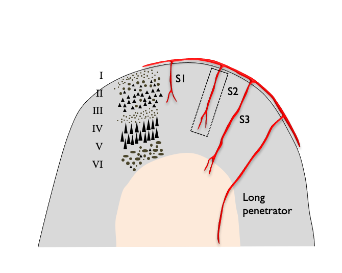
To break this down, let's see how to design only a part of the brain 🧠: a piece of the cortex and some small vessels
#MedStudentTwitter #MedEd #FOAMed #tweetorial #FOAMrad #Neurology #Neurosurgery #RadEd #illustration #PowerPoint
3/
#MedStudentTwitter #MedEd #FOAMed #tweetorial #FOAMrad #Neurology #Neurosurgery #RadEd #illustration #PowerPoint
3/

👉🏿This illustration is in fact comprised of 4 individual elements (some are groups) that are created separately, but with the final piece in mind.
(I usually sketch🖌️ on paper w pencils the basic drawing that I aim to create, to get the general idea)
4/
(I usually sketch🖌️ on paper w pencils the basic drawing that I aim to create, to get the general idea)
4/

Step 1: Curve tool - gives enormous control for drawing the basic shapes you need
➰Now, I have created the basic shape for the cortex 👇🏿
I will do a similar, but smaller shape for the white matter...
5/

➰Now, I have created the basic shape for the cortex 👇🏿
I will do a similar, but smaller shape for the white matter...
5/


Step 2: add color🎨("Shape fill" tool) and outline
(you can do fancy things like gradient fills, shadows, 3D, but let's go with a simple fill)
Step 3: layering🃏- you overlay shapes to create the whole
Now a basic piece of cortex and some white matter is taking shape..👇🏿
6/
(you can do fancy things like gradient fills, shadows, 3D, but let's go with a simple fill)
Step 3: layering🃏- you overlay shapes to create the whole
Now a basic piece of cortex and some white matter is taking shape..👇🏿
6/

Step 4: Correction of details with the "Edit points" tool under magnification
**each shape is comprised of individual connected points, you just don't normally see them!
I use this to make small alterations to shapes: change curvature of points, angle, add/delete points...
7/

**each shape is comprised of individual connected points, you just don't normally see them!
I use this to make small alterations to shapes: change curvature of points, angle, add/delete points...
7/


Step 5: To create the blood vessel, I use again the Curve tool under magnification to make the outline of the shape, in this case a penetrating arteriole.
... add color, outline, edit points PRN
See for example a smaller arteriole in the making👇🏿
(you can sprinkle VEGF)
8/


... add color, outline, edit points PRN
See for example a smaller arteriole in the making👇🏿
(you can sprinkle VEGF)
8/


As you would imagine, the more elaborate a shape is, the more painstaking the process of modifying the points is...
9/
9/

In some illustrations or parts of them, if I aim for #Anatomy #neuropath #NeuroRad #biological accuracy, I might consult an atlas or an actual photo.
This is what I used for example to design the #cortical layers 👇🏿
10/

This is what I used for example to design the #cortical layers 👇🏿
10/


⚫️ Now, just for fun, let's add some @microbleeds in the cortex.
I draw a circe, paint it black, add a bit of black glow to represent the blooming effect on blood-sensitive MRI, make copies, and add them to the illustration 👇🏿
11/

I draw a circe, paint it black, add a bit of black glow to represent the blooming effect on blood-sensitive MRI, make copies, and add them to the illustration 👇🏿
11/


🧠🏗️ And this is how you build a brain, piece (of cortex) by piece...
I use more or less the same method for more complicated illustrations.
I hope you find this helpful!
I use more or less the same method for more complicated illustrations.
I hope you find this helpful!

🤿More illustrations in this "Moment"
https://twitter.com/a_charidimou/status/1530913839138709509?s=20&t=RzIInVoQLQqI_ZPZHbh_9w
• • •
Missing some Tweet in this thread? You can try to
force a refresh


