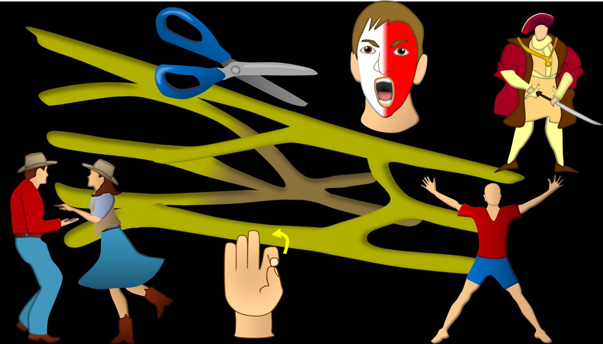
1/Ready for some heavy lifting? My second #tweetorial on the BRACHIAL PLEXUS! This time we cover how the #brachialplexus looks on #MRI.

#medtwitter #meded #neurosurgery #orthotwitter #orthopedics #neurorad #radres #medstudent #FOAMed #FOAMrad #spine #radiology #neurotwitter Image
2/Brachial plexus is how the cervical nerves reach the arm. In the coronal plane, it looks like a slide, guiding nerves downward. Bc nerves are traveling laterally, sagittal MRI plane is our plane of choice to cut the nerves in cross section & see down the barrel of the nerves Image
3/But it’s more than a slide, it’s a complex highway, w/nerves joining & dividing—like highway off ramps & on ramps. If you want to know more about this intrinsic anatomy, see my first brachial plexus tweetorial here: Image
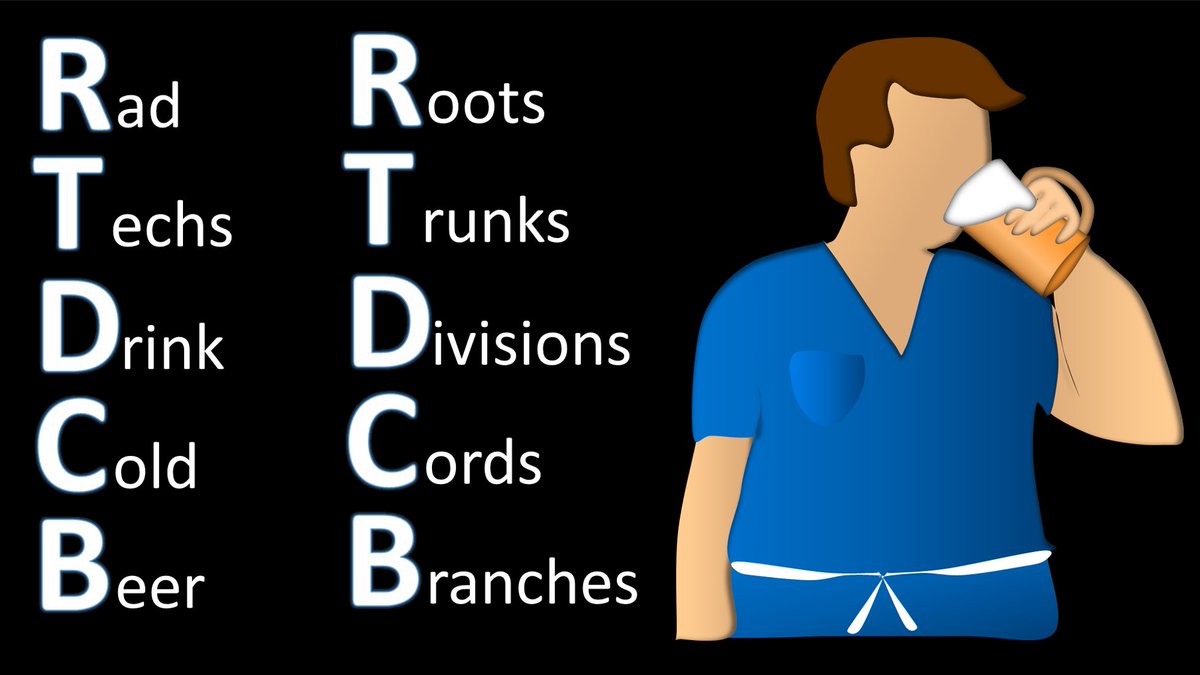
4/The most medial sagittal cut, right after the neural foramina, shows us the roots (Remember Rad Techs Drink Cold Beer). In the sagittal plane, the roots look like the rungs of a ladder. This makes sense, bc we are climbing up the ladder of the slide to go down to the arm Image
5/The anatomic landmark for the roots is the 1st rib head. I remember this bc the roots are closest to the CNS or HEAD, so they are by the rib HEAD. The roots together w/the rib head make the ladder look like a folding ladder, w/the rib head supporting the ladder root rungs Image
6/Here are the roots on a sagittal MRI—ladder rungs are the roots, supported like a folding ladder by the first rib head. Image
7/Next are the trunks. Trunks on sagittal MRI are easy—they are arranged like, wait for it…a tree TRUNK. They are right behind the subclavian artery, which looks like a little shrub in front of the tree trunk. Image
8/There are 2 anatomic landmarks for the trunks. Trunks are at the posterior 1st rib & in between the scalene muscles. The rib & the scalene look like an A-frame house, one that people often have for cabins in the woods. So you have trees & bushes in front of a A-frame cabin. Image
9/Here are the trunks on a sagittal MRI, with the trunks looking like, well, trunks & the scalene muscles making the A-frame cabin in the woods in the background. Image
10/Next is the divisions. I remember what these look like on sagittal images by remembering that DIVISIONS are DIVINE /DRESSY—all w/the letter D. They look like a fancy hair updo on top of the subclavian artery head. Image
11/The anatomic landmark for the divisions is the clavicle. The divisions sit BEHIND the clavicle. I remember this bc Victorian ladies with fancy updos will have fans that they hide their face behind. Similarly, the divisions hide behind the clavicle. Image
12/Here are the divisions on a sagittal MRI, they are clumped together like a fancy bun above the head of the subclavian. The clavicle is in front of them, allowing them to keep their Victorian modesty from the prying eyes of men Image
13/Next are the cords. The subclavian artery and the cords are organized so they look like a paw print. I remember this bc both Cord and Cat start w/C. So cords make a cat claw print. Image
14/Anatomic landmark for the cords is the coracoid. Cords are underneath the coracoid. I remember this bc Cats who make the paw prints are always hiding under something like a couch. So the cords hide under the coracoid. I remember it’s the CORacoid bc cats hide when CORnered Image
15/Here are the cords on a sagittal MRI, with the paw print hiding underneath the coracoid process above it. Image
16/Last are the branches. In the sagittal plane, the subclavian artery together w/the branches looks like a fat beetle with four legs. I remember this bc Branches and Beetle, or Bug, all start w/B. Image
17/Here are the branches on sagittal MRI, w/the subclavian artery as fat body of the beetle and the branches as the beetle’s arms. Image
18/You can remember this w/an old fashioned fairy tale--about a house in the woods, home to a divine but shy princess. She had a cat that hid under things & ate all bugs in the home, bc no princess wants bugs! Now you know the imaging anatomy. Next tweetorial will be pathology! Image

• • •

Missing some Tweet in this thread? You can try to force a refresh
 

Keep Current with Lea Alhilali, MD

Lea Alhilali, MD Profile picture

Stay in touch and get notified when new unrolls are available from this author!

Read all threads

This Thread may be Removed Anytime!

PDF

Twitter may remove this content at anytime! Save it as PDF for later use!

Try unrolling a thread yourself!

how to unroll video
  1. Follow @ThreadReaderApp to mention us!

  2. From a Twitter thread mention us with a keyword "unroll"
@threadreaderapp unroll

Practice here first or read more on our help page!

More from @teachplaygrub

Sep 30
1/Raise your hand if you’re confused by the BRACHIAL PLEXUS! I could never quite remember or understand it—but now I do & I’ll show you how
A #tweetorial on #brachialplexus #anatomy!
#medtwitter #meded #neurosurgery #orthotwitter #orthopedics #neurorad #radres #medstudent #FOAMed Image
2/Everyone has a mnemonic to remember brachial plexus anatomy. I’m a radiologist, so I remember one about Rad Techs. But just remembering the names & their order isn’t enough. That is just the starting point--let’s really understand it Image
3/From the mnemonic, we start with the roots—the cervical nerve roots. I remember which roots make up the brachial plexus by remembering that it supplies the hand. You have 5 fingers on your hand so we start with C5 & we take 5 nerve roots (C5-T1). Image
Read 21 tweets
Sep 27
1/ Talk about dangerous liaisons! Abnormal brain vascular connections can be dangerous
A #tweetorial about an important abnormal connection: dural arteriovenous fistulas (dural AVF) in collaboration w/ @SVINJournal!

Featuring this 🆓#openaccess article: ahajournals.org/doi/10.1161/SV…
2/Dural sinuses sit inside dural leaflets. Arteries that feed the dura also feed the walls of sinuses, like vasa vasorum. Arteries in the walls of veins are a natural connection between the veins and arteries—but these connections are usually closed in normal pts.
3/Whether these connections are open depends on pressure. Like a hose w/a hole in it, at normal pressures, abnormal connections are not open. But if pressure is increased w/thrombosis or stenosis, the connections open, like high pressure water squirting out through a hole.
Read 16 tweets
Sep 25
1/Sometimes the tiniest thing can be the biggest pain—that’s microvascular compression of the trigeminal nerve! But seeing such a tiny finding can be hard!

Here’s a 🧵 about how to look at the trigeminal nerve on MRI. #medtwitter #neurotwitter #neurorad #meded #FOAMed #HNrad Image
2/The most important thing to remember is that the nerve is 3D so you have to look for compression in all 3 planes. Let’s start w/the axial plane. On a normal axial, the trigeminal nerves should look like the arms of an alien sticking out of the pons. Image
3/Compression in the axial plane usually will deviate the nerve laterally—making it so that the Alien looks like he is flexing one of his arms. So if you see the Alien trying to show his guns—that’s microvascular compression! Image
Read 8 tweets
Sep 19
1/Need help reading spine imaging? I’ve got your back!
A #tweetorial about the ABCs of reading spine MRs & CTs.
#medtwitter #FOAMed #FOAMrad #medstudenttwitter #medstudent #neurorad #radres #neurosurgery #spine #orthopedics @MedTweetorials @StefanTigges Image
2/A is for alignment. Normal spinal alignment is perfectly in balance, resulting in the minimal energy needed for erect posture. Even subtle changes in alignment need compensatory changes to maintain posture, resulting in more work/energy expenditure & pain. Image
3/The goals for alignment on imaging: (1) look for unstable injuries & (2) look for malalignment that causes early degenerative change. Abnormal motion causes spinal elements to abnormally move against each other, like grinding teeth wears down teeth—this wears down the spine Image
Read 19 tweets
Sep 10
1/Don't fall for the siren song of calling all bright round objects at foramen of Monro colloid cysts. Like a true siren song, this may be a trap!
A🧵about lesions in this region that can trap you
#medtwitter #FOAMed #FOAMrad #medstudent #neurorad #radres #meded #Neurosurgery
2/Here are 3 lesions, all round and bright and in the region of the foramen of Monro. Can you tell from the images which is a colloid cyst and which may be something else? Choose which one or ones you think are a colloid cyst
Choose which one you think is a colloid cyst
Read 13 tweets
Sep 8
1/Controversy in radiology gets tense! The Mt Fuji sign for tension pneumocephalus is under scrutiny.When should you call it?
A #tweetorial about #imaging this important #neurosurgery complication
#medtwitter #FOAMed #FOAMrad #medstudent #neurorad #radres #meded #neurotwitter Image
2/Some believe that the peaked, mountain like appearance of the frontal lobes is a critical sign of a life-threatening complication & should be called & reported. Others believe it is too non-specific, is commonly seen when there isn’t tension & should be retired. Who’s right? Image
3/First, let’s clarify about what the Mt Fuji sign actually is. Most are familiar with the fact that large collections of pneumocephalus can compress the frontal lobes—making them look like the slopes of a mountain. But this isn’t actually enough to call Mt Fuji. Image
Read 12 tweets

Did Thread Reader help you today?

Support us! We are indie developers!


This site is made by just two indie developers on a laptop doing marketing, support and development! Read more about the story.

Become a Premium Member ($3/month or $30/year) and get exclusive features!

Become Premium

Don't want to be a Premium member but still want to support us?

Make a small donation by buying us coffee ($5) or help with server cost ($10)

Donate via Paypal

Or Donate anonymously using crypto!

Ethereum

0xfe58350B80634f60Fa6Dc149a72b4DFbc17D341E copy

Bitcoin

3ATGMxNzCUFzxpMCHL5sWSt4DVtS8UqXpi copy

Thank you for your support!

Follow Us on Twitter!

:(