A 29-YO♂️, 6 months before, HIV + & non–drug-resistant pulmonary tuberculosis, antiretroviral & 4-drug antituberculous therapy initiated but soon reduced to rifampin & isoniazid only: abdominal pain on the L side
CT: ?
1/5

DOI: 10.1056/NEJMicm2206174
#radiologist #GITwitter
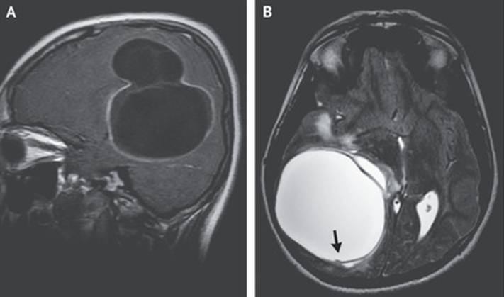
CT: an enlarged spleen with numerous hypodense lesions (A).
CD4 cell count: 119/mm3
VIH viral load: 1778 copies/mm3

A splenectomy was performed to evaluate for cancer:
numerous necrotic nodules with purulent discharge (B).
2/5

#microbiology #gastroenterology #IDtwitter
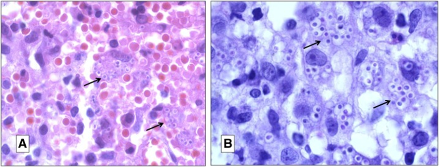
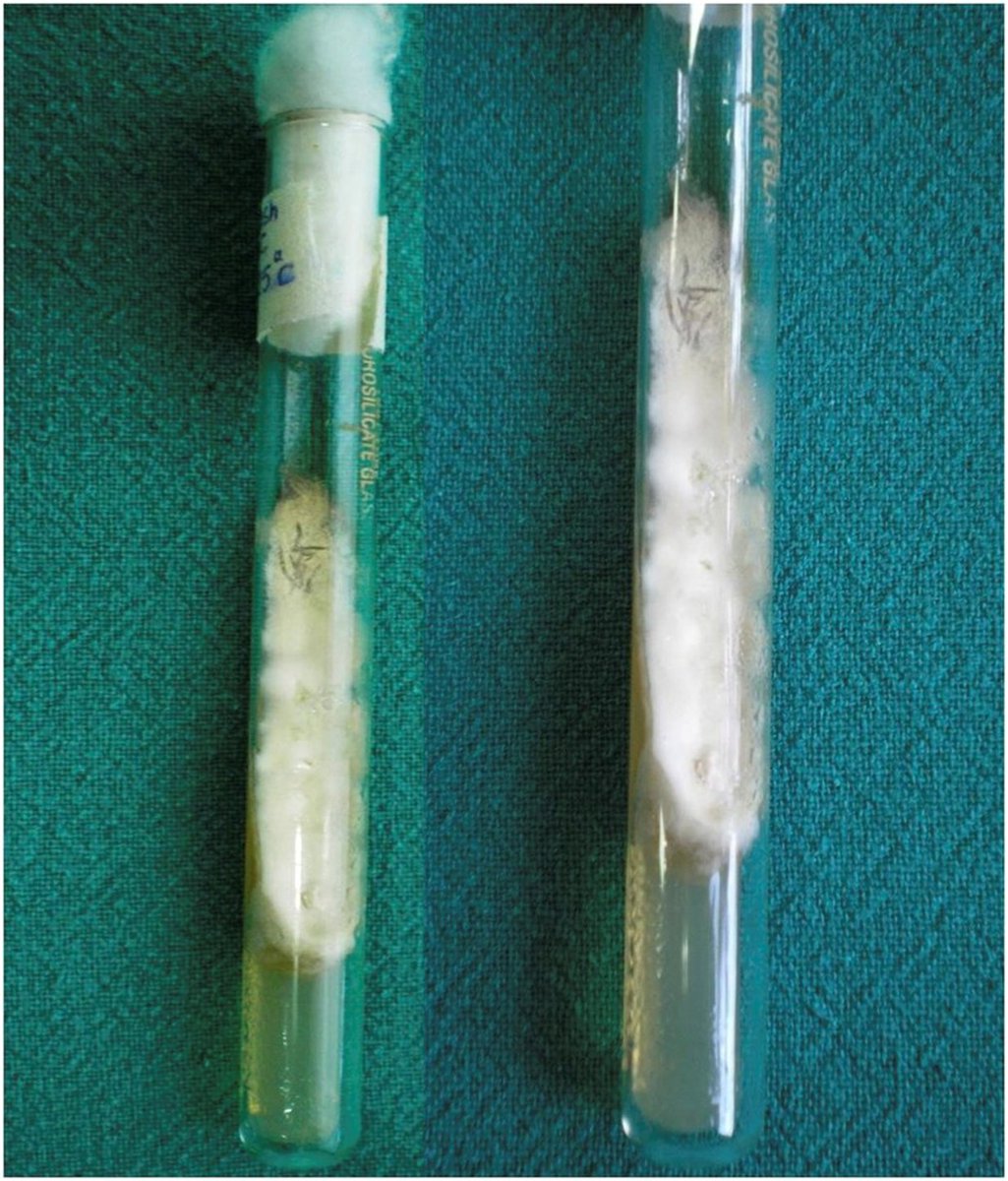
🔬granulomatous inflammation with caseous necrosis (C) and acid-fast bacilli (D, arrowheads).
A tissue🧫: ➖

PCR: ➕for Mycobacterium tuberculosis

SPLENIC TUBERCULOSIS
3/5

#bacteriology #MedTwitter #GIPath
Pyrazinamide and ethambutol were added back to the patient’s antituberculous regimen

The patient completed 9 months of four-drug therapy.
4/5

#Doctor #MedStudentTwitter #resident
Splenic tuberculosis:
📌a rare clinical manifestation of tuberculosis
📌typically described in immunocompromised patients
📌usually seen in association with disseminated miliary tuberculosis
5/5

DOI: 10.1056/NEJMicm2206174
#medicine #internalmedicine #primarycare

• • •

Missing some Tweet in this thread? You can try to force a refresh
 

Keep Current with Antonio Guerrero

Antonio Guerrero Profile picture

Stay in touch and get notified when new unrolls are available from this author!

Read all threads

This Thread may be Removed Anytime!

PDF

Twitter may remove this content at anytime! Save it as PDF for later use!

Try unrolling a thread yourself!

how to unroll video
  1. Follow @ThreadReaderApp to mention us!

  2. From a Twitter thread mention us with a keyword "unroll"
@threadreaderapp unroll

Practice here first or read more on our help page!

More from @guerreroespejo

Feb 10
A 17-YO: a 3-week of dizziness, headache, & weakness of the R leg

MR: ring-enhancing lesions in the frontal lobe & basal ganglia on the L side (A; T1-W) as well as surrounding edema & midline shift (B; T2-W)
1/6

DOI: 10.1056/NEJMicm2202196
#neurology #radiology #IDTwitter
IgG ab for Echinococcus multilocularis & E. granulosus: ➖

Surgical excision🔬: necrotic tissue, granulomatous inflammation, echinococcal laminated membrane (C, arrow) & an intact cyst (D, arrow)

RT-PCR: ➕for E. multilocularis
2/6

#parasitology #Pathologists #pathology
CT imaging of the chest and abdomen did not show any other sites of disease.

CEREBRAL ALVEOLAR ECHINOCOCCOSIS
3/6

DOI: 10.1056/NEJMicm2202196
#microbiology #MedTwitter #Doctor
Read 6 tweets
Feb 9
A 14-yo ♂️ lived on a farm: a 1-month history of episodic headaches, vomiting, & papilledema
MR: a multiloculated cyst of the brain (A) with a hypointense rim and small projections in T2 phase (B, arrow)
1/5

DOI: 10.1056/NEJMicm2208104
#neurology #radiology #pediatric Image
Findings suggestive of CYSTIC ECHINOCOCCOSIS.

CT of the body: no other sites of disease.
A craniotomy was performed, during which saline irrigation was used to separate the cyst wall from the brain to avoid rupture.
2/5

DOI: 10.1056/NEJMicm2208104
#IDtwitter #parasitology Image
🔬: an echinococcal laminated membrane lined by a germinal layer with daughter cysts (Panel D, arrows) and protoscolices (inset, arrows) with hooklets (arrowhead).

PRIMARY CEREBRAL CYSTIC ECHINOCOCCOSIS FROM ECHINOCOCCUS GRANULOSUS
3/5

#microbiology #Pathologists #pathology Image
Read 5 tweets
Feb 7
A 50-YO♂️of North India, visited bat caves & a bird sanctuary in Thailand: multiple plaques & nodules on his face, neck, arms & trunk with hoarseness, fever, weight loss for months
Laryngoscopy: nodules over the tonsils & posterior pharyngeal wall
1/4

#medicine #dermatology
Skin & laryngeal 🔬: histiocytes with intracellular & extracellular PAS+ yeast cells
Skin biopsy🧫: tuberculate macroconidia confirmed as H. capsulatum var. capsulatum
Ab against Histoplasma by ID:➕

DISSEMINATED HISTOPLASMOSIS
2/4

doi.org/10.3109/136937…
#mycology #IDtwitter
Disseminated histoplasmosis involving skin & larynx in an apparently immunocompetent individual

The infection appeared to be imported from Thailand.

The patient responded very well to intravenous amphotericin B followed by itraconazole.
3/4

#microbiology #MedTwitter #Doctor
Read 4 tweets
Feb 1
A 46-YO ♂️: oropharyngeal lesions, cervical lymphadenopathy, intermittent fever, hepatosplenomegaly, and deteriorating general health.

Needle aspiration from submandibular lymph node: intra- and extracellular yeasts
1/4

DOI:
doi.org/10.4269/ajtmh.…
#dermpath #mycology Mucocutaneous examination showed multiple well defined, disc
The histopathology of oral lesion showed features of HISTOPLASMOSIS confirmed later from culture of Histoplasma capsulatum on culture
2/4

#IDtwitter #microbiology #MedTwitter Fine needle aspiration cytology from submandibular lymph nod
In immunocompetent hosts, when chronic disseminated histoplasmosis occurs from pulmonary infection, oral ulcers, nodules, and vegetative plaques are common and dissemination to liver, spleen, bone marrow, and other organs is frequent.
3/4

#Doctor #MedStudentTwitter #resident
Read 4 tweets
Jan 31
A 37-YO, farmer in India, #HIV+ immunecompromised ♂️, treatment for suspected pulmonary tuberculosis: papulonodular lesions of 3-month duration over the face, neck, trunk, limbs, palms & soles, & oropharynx; fever & generalized lymphadenopathy
🔬: ?
1/7

#IDTwitter #microbiology Wide spread papules and nodules over face. Similar lesions w
🔬: Blastoconidias within the macrophages.
🧫: Histoplama capsulatum: characteristic colony, conversion to yeast form, characteristic🔬appearance.

PROGRESSIVE DISSEMINATED HISTOPLASMOSIS
2/7

doi: 10.4269/ajtmh.17-0432
#dermpath #mycology #internalmedicine 1.- Histologic section of skin lesion showing sheets of macr
Diagnosis is usually suspected on visualization of characteristic intracellular yeast (blastoconidias), the pathogenic or tisular form, with a clear rim and garnulomatous cellular infiltrate in tissue specimen or lymph node aspirate
3/7

#MedTwitter #Doctor
Read 7 tweets
Jan 30
39 años ♂️: hiperqueratosis subungueal y coloración amarillo-verdosa de la lámina ungueal del 5º dedo de pie dcho. de 2 años de evolución.

1/ 7

✍️B. Monteagudo, O. Figueroa, @olaya_sm, S. Méndez
DOI: 10.1016/j.ad.2018.04.013
#dermatología #microbiología
@olaya_sm Con 🔍: Hiperqueratosis subungueal (&), borde distal irregular, zonas parduzcas de aspecto hemorrágico (&) y patrón multicolor de lámina ungueal con áreas verdes ($) y azul-negruzcas (*), y otras longitudinales amarillentas (→)

CLORONIQUIA
2/7

#dermatology
@olaya_sm 🧫 uña: Pseudomonas aeruginosa y hongo filamentoso (imposible identificar por sobrecrecimiento bacteriano).

UÑA VERDE CAUSADA POR ONICOMICOSIS COINFECTADA POR PSEUDOMONAS AERUGINOSA

Con ciprofloxacino oral y terbinafina, tres meses: resolución.
3/7

#AtencionPrimaria #micología
Read 6 tweets

Did Thread Reader help you today?

Support us! We are indie developers!


This site is made by just two indie developers on a laptop doing marketing, support and development! Read more about the story.

Become a Premium Member ($3/month or $30/year) and get exclusive features!

Become Premium

Don't want to be a Premium member but still want to support us?

Make a small donation by buying us coffee ($5) or help with server cost ($10)

Donate via Paypal

Or Donate anonymously using crypto!

Ethereum

0xfe58350B80634f60Fa6Dc149a72b4DFbc17D341E copy

Bitcoin

3ATGMxNzCUFzxpMCHL5sWSt4DVtS8UqXpi copy

Thank you for your support!

Follow Us on Twitter!

:(