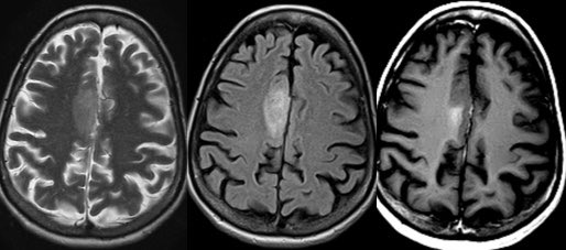
Case of a radiation induced pseudoaneurysm in this patient with headache and AMS 🧠
Imaging in thread #Neurosurgery #Neurology #neurotwitter #radres #MedEd #MedTwitter @TheASNR


Imaging in thread #Neurosurgery #Neurology #neurotwitter #radres #MedEd #MedTwitter @TheASNR


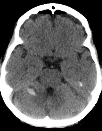
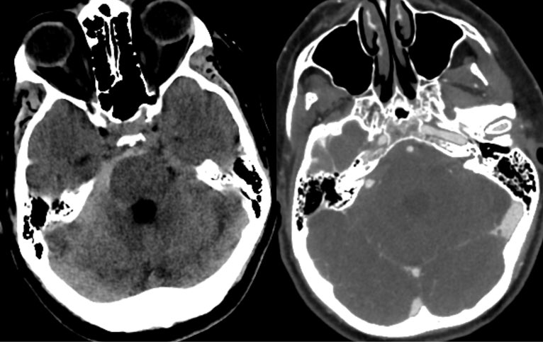
▶️Initial head CT shows subarachnoid hemorrhage centered in the right cerebellopontine angle cistern
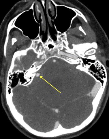
▶️CTA confirms an aneurysm of the right anterior inferior cerebellar artery (AICA)

▶️CTA confirms an aneurysm of the right anterior inferior cerebellar artery (AICA)

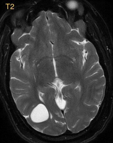
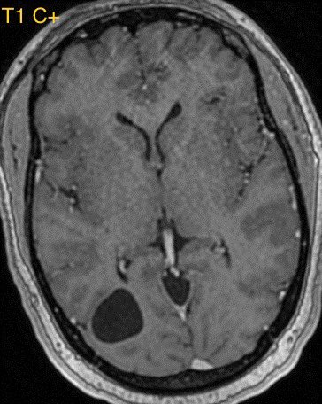
▶️MR displays and ice cream shaped enhancing mass extending through the right internal auditory canal into the cerebellopontine angle cistern, consistent with a vestibular schwannoma #icecream
▶️Careful search into the history confirms the schwannoma was treated with radiation

▶️Careful search into the history confirms the schwannoma was treated with radiation


Learning points:
💡 The Anterior inferior cerebellar artery (AICA) normally crosses the 8th cranial nerve at the CP angle cistern
💡When masses are treated with radiation, they can occasionally induced pseudoaneurysms of nearby vessels which are high risk for rupture

💡 The Anterior inferior cerebellar artery (AICA) normally crosses the 8th cranial nerve at the CP angle cistern
💡When masses are treated with radiation, they can occasionally induced pseudoaneurysms of nearby vessels which are high risk for rupture

• • •
Missing some Tweet in this thread? You can try to
force a refresh
 Read on Twitter


