Daniel Gewolb, MD Profile picture
May 19 9 tweets 5 min read Twitter logo Read on Twitter
Interesting case of complicated acute bacterial rhinosinusitis in this child with no PMH presenting w/ HA, fever & L sided weakness

#NeuroTwitter #ent #radres #neurosurgery @TheASNR @ASHNRSociety @PhilipRChapman1 #radres #futureradres ImageImageImageImage
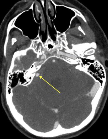
CT shows opacification of the frontal and anterior ethmoid sinuses without evidence of cortical dehiscence

💡 In peds, infection can spread through vascular channels w/o destroying the bone ImageImage
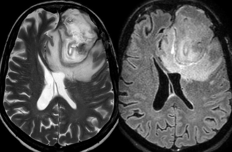
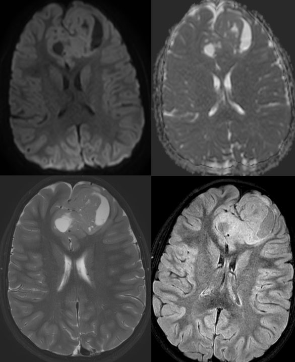
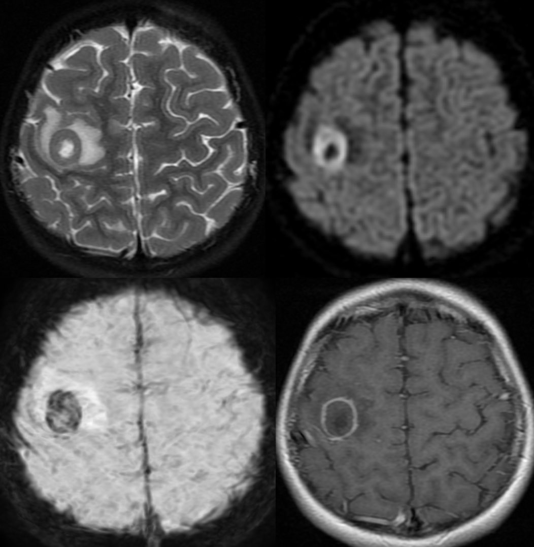
MR shows abnormal signal filling the sinuses with associated restricted diffusion 2/2 purulent material

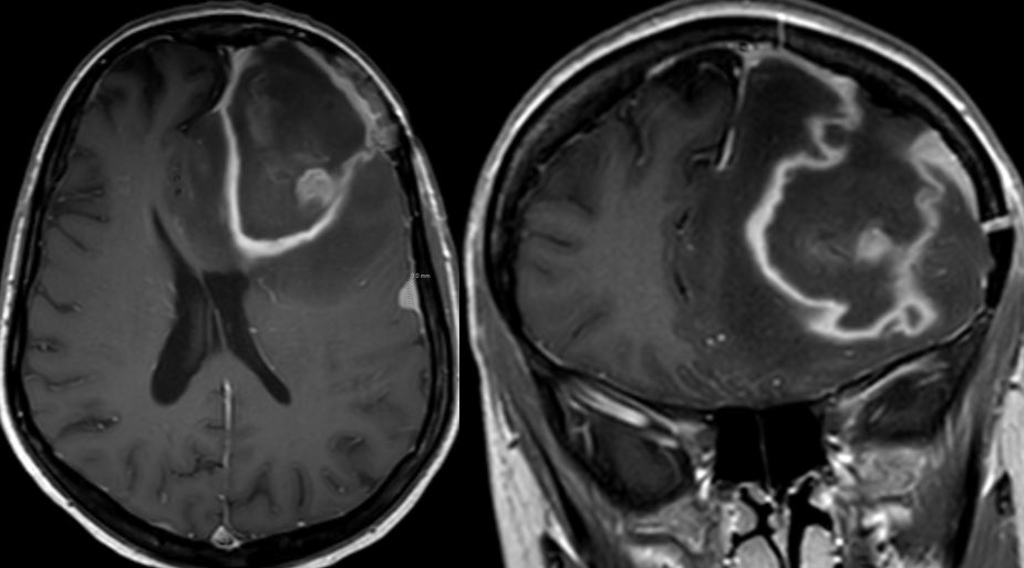
Post contrast we see areas of relative hypoenhancement in comparison to the normal mucosa on the contralateral side ImageImageImage
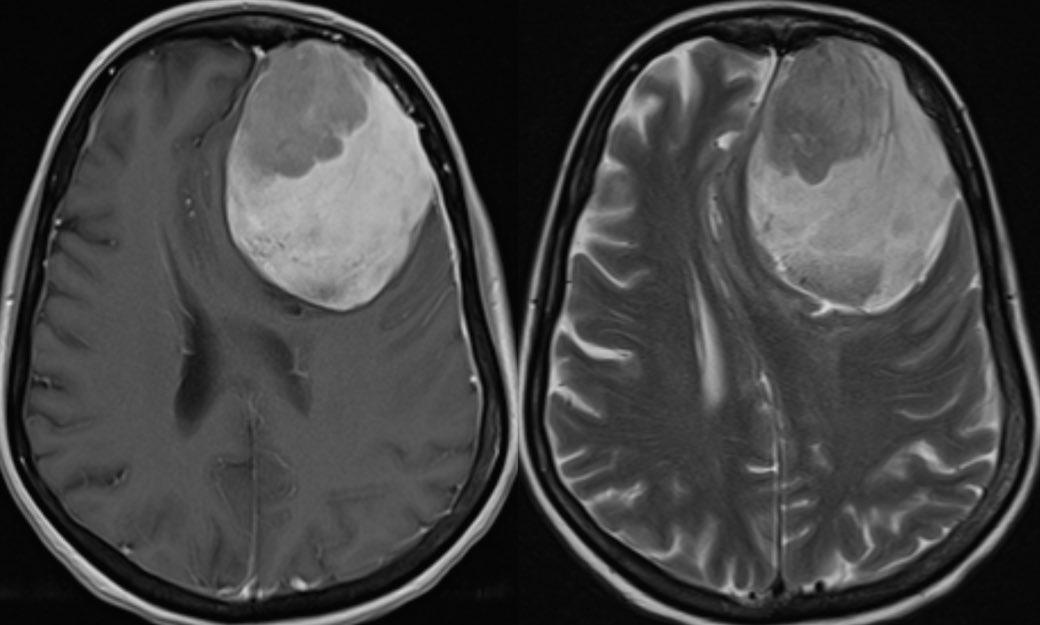
There is smooth pachymeningeal enhancement, suggestive of intracranial extension as well as an abscess in the right frontal lobe ImageImage
Learning points:

💡 Acute sinusitis is a clinical diagnosis (imaging has poor correlation w/ symptoms as there is a high incidence of mucosal abnormalities in asymptomatic patients)

💡 Even normal sinus CT can have endoscopic evidence of sinusitis
💡 imaging can be used when complications are suspected

💡 CT sinuses can also be used in the outpatient setting in patients with chronic or recurrent sinusitis as a preoperative evaluation in those who may benefit from FESS
💡 Reduced or absent mucosal enhancement is NOT pathognomonic for invasive fungal sinusitis as it can be seen in mucoceles (possibly with superimposed infection) and patchy asymmetric diminished enhancement in bacterial sinusitis
Possible mechanism is increased sinus pressure leading to reduced perfusion or possible mucosal necrosis (if it can invade bone why not cause mucosal necrosis)? This is just speculation, I do not know what actually occurs 🤷🏻‍♂️
💡 Invasive fungal is usually thickened absent mucosal enhancement, no restricted diffusion, bone erosions and immunocompromised host

• • •

Missing some Tweet in this thread? You can try to force a refresh
 

Keep Current with Daniel Gewolb, MD

Daniel Gewolb, MD Profile picture

Stay in touch and get notified when new unrolls are available from this author!

Read all threads

This Thread may be Removed Anytime!

PDF

Twitter may remove this content at anytime! Save it as PDF for later use!

Try unrolling a thread yourself!

how to unroll video
  1. Follow @ThreadReaderApp to mention us!

  2. From a Twitter thread mention us with a keyword "unroll"
@threadreaderapp unroll

Practice here first or read more on our help page!

More from @daniel_gewolb

May 21
Learning case in this 40 y/o F with history of whole brain radiation as a child for brain tumor treatment
#NeuroRad #neurosurgery #Neurology @TheASNR #NeuroTwitter #meded #radres ImageImageImageImage
Brain radiation is a risk factor for the development of meningiomas …this patient subsequently developed multiple meningiomas including this large frontal meningioma Image
The mass was treated and immediate post op CT shows expected post op change with the resection cavity, some hemorrhage, and pneumocephalus Image
Read 9 tweets
Mar 27
What is the most likely diagnosis in this adolescent with seizure? 🧠

(Sorry I have no CT without)

#neurotwitter #peds #Neurosurgery #Neurology @The_ASPNR @TheASNR #MedTwitter
What is the most likely diagnosis?
Answer: Confirmed supratentorial ependymoma

Predicting tumors is incredibly challenging in the absence of specific features …some learning points on the case in 🧵
Read 8 tweets
Mar 27
Case of a radiation induced pseudoaneurysm in this patient with headache and AMS 🧠

Imaging in thread #Neurosurgery #Neurology #neurotwitter #radres #MedEd #MedTwitter @TheASNR ImageImageImageImage
▶️Initial head CT shows subarachnoid hemorrhage centered in the right cerebellopontine angle cistern

▶️CTA confirms an aneurysm of the right anterior inferior cerebellar artery (AICA) ImageImage
▶️MR displays and ice cream shaped enhancing mass extending through the right internal auditory canal into the cerebellopontine angle cistern, consistent with a vestibular schwannoma #icecream

▶️Careful search into the history confirms the schwannoma was treated with radiation ImageImage
Read 4 tweets
Mar 21
What is the most likely diagnosis in this 25 y/o M with headache? 🧠

Answer later tonight #radres #Neurology #Neurosurgery #MedEd #MedTwitter #NeuroTwitter @RSNA ImageImageImageImage
Most likely diagnosis?
Answer: confirmed germinoma, all these masses are on the differential for a pineal region mass …perhaps the most helpful clue is the age and gender rather than the imaging 🧠
Read 7 tweets
Mar 20
Glioblastoma is the most common variety of astrocytoma

The presence of necrosis is the characteristic feature of glioblastoma

Imaging details in thread #Neurosurgery #neurotwitter #radres #MedTwitter #Neurology @TheASNR
Some correlates for high grade astrocytoma include necrosis, marked mass effect, restricted diffusion (often patchy, eccentric or nodular in glioblastoma), elevated CBV, enhancement, and older age

The presence of ependymal spread is another poor prognostic sign (shown below)
Important learning point: when dealing with glioblastoma, the neoplasm extent is not defined by the area of enhancement but rather by the entire area of T2/FLAIR abnormality. Microscopic infiltrating tumor clearly extends beyond the areas of enhancement. 🧠
Read 5 tweets
Mar 16
Preoperative approach to sellar region masses, what the surgeon needs to know (at least what I think they need to know)

Additional reporting tips from surgeons are welcomed and encouraged! #Neurosurgery @TheASNR #radres #MedEd #MedTwitter #futureradres #endocrine #Neurology
1️⃣Where is the mass located?

Is it sellar based? suprasellar (S)? clival? Planum sphenoidale (PS)? Tuberculum sellae (arrow)?

▶️Location can change operative approach including but not limited to subfrontal vs transsphenoidal
2️⃣Where is the normal pituitary gland?

▶️this can be tough when the mass is large but the normal tissue often enhances more avidly than tumor so look for a strip of relatively avid enhancement along the periphery of the mass. Surgeons do not want to remove normal pit tissue
Read 10 tweets

Did Thread Reader help you today?

Support us! We are indie developers!


This site is made by just two indie developers on a laptop doing marketing, support and development! Read more about the story.

Become a Premium Member ($3/month or $30/year) and get exclusive features!

Become Premium

Don't want to be a Premium member but still want to support us?

Make a small donation by buying us coffee ($5) or help with server cost ($10)

Donate via Paypal

Or Donate anonymously using crypto!

Ethereum

0xfe58350B80634f60Fa6Dc149a72b4DFbc17D341E copy

Bitcoin

3ATGMxNzCUFzxpMCHL5sWSt4DVtS8UqXpi copy

Thank you for your support!

Follow Us on Twitter!

:(