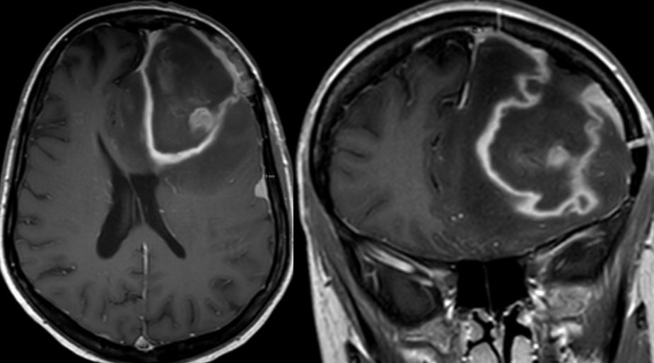
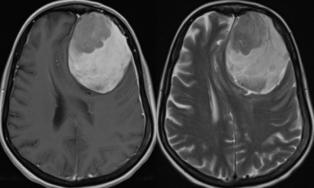
Child with a history of dental caries presents with a firm mass at the angle of the mandible. What is the most likely diagnosis? 🤔 🧠
#neurotwitter #ent #peds #Neurology #neurosurgery @ASHNRSociety @The_ASPNR #MedTwitter


#neurotwitter #ent #peds #Neurology #neurosurgery @ASHNRSociety @The_ASPNR #MedTwitter


Answer: Sclerosing osteomyelitis of Garré
▶️Biopsy showed a reactive and reparative osseous process and bone culture grew oral flora (though cultures are usually negative)
▶️Biopsy showed a reactive and reparative osseous process and bone culture grew oral flora (though cultures are usually negative)
▶️SOG is thought to be due to a low grade infection possibly 2/2 dental disease. However, there should be no signs of acute infection (suppuration, bony sequestration or draining tracts)
▶️SOG is considered a variant of chronic non-suppurative osteomyelitis and is more common in male children
▶️CRMO and SAPHO may be related to or the same diseases and are indistinguishable to my knowledge (would love input from those more experienced here)
▶️CRMO and SAPHO may be related to or the same diseases and are indistinguishable to my knowledge (would love input from those more experienced here)
Imaging:
▶️Unilateral (usually) mandibular involvement
▶️Dense CORTICAL thickening
▶️Onion skin PERIOSTEAL reaction (aka periostitis Ossificans)
▶️Narrowed medullary cavity
▶️Unilateral (usually) mandibular involvement
▶️Dense CORTICAL thickening
▶️Onion skin PERIOSTEAL reaction (aka periostitis Ossificans)
▶️Narrowed medullary cavity
▶️Fibrous dysplasia is the main ddx which has the ground glass matrix but EXPANDS the medullary cavity and THINS the cortex with NO periosteal reaction. FD is also more common in the maxilla than the mandible
▶️Biopsy often necessary to exclude malignant processes such as osteosarcoma
Ddx for sclerotic dental lesions:
Odontoma
Cementoosseous dysplasia
Fibrous dysplasia
Sclerosing osteomyelitis of Garré
Sarcoma (ewings, osteo, chondro)
Mets
Cementoblastoma
Padgets
Lymphoma


Ddx for sclerotic dental lesions:
Odontoma
Cementoosseous dysplasia
Fibrous dysplasia
Sclerosing osteomyelitis of Garré
Sarcoma (ewings, osteo, chondro)
Mets
Cementoblastoma
Padgets
Lymphoma


• • •
Missing some Tweet in this thread? You can try to
force a refresh

 Read on Twitter


