Is anyone planning to do some reading about hippocampal neuroanatomy over the holidays?
If you answered, "yes", this week's #SubfieldWednesday is for you! We will give you a list of "must read" atlas references about our favorite brain structure. 🍤❣️
#SubfieldWednesday (1/n)
If you answered, "yes", this week's #SubfieldWednesday is for you! We will give you a list of "must read" atlas references about our favorite brain structure. 🍤❣️
#SubfieldWednesday (1/n)
In our 2015 paper (Yushkevich et al., NeuroImage, 2015), we provided a list of common atlases used for hippocampal subfield definition across labs.
#SubfieldWednesday (2/n)
#SubfieldWednesday (2/n)

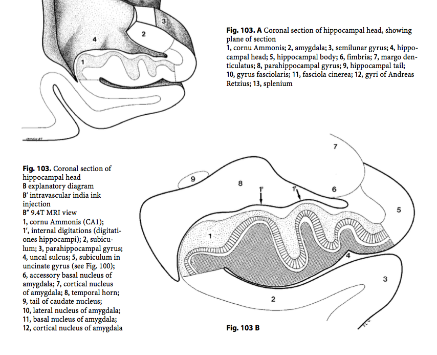
One of the most commonly used atlases cited was:
Duvernoy, H. M. (2005). The human hippocampus: functional anatomy, vascularization and serial sections with MRI. Springer Science & Business Media.
#SubfieldWednesday (3/n)

Duvernoy, H. M. (2005). The human hippocampus: functional anatomy, vascularization and serial sections with MRI. Springer Science & Business Media.
#SubfieldWednesday (3/n)


Duvernoy's Chapter 7: "Sectional Anatomy and Magnetic Resonance Imaging" is especially helpful for helping the reader visualize the subfields on MRI and post-mortem sections.
#SubfieldWednesday (4/n)


#SubfieldWednesday (4/n)


Insausti & Amaral's, "The Hippocampal Formation" is another commonly cited atlas. The definitions in Figure 23.15 influenced a number of existing MRI protocols.
Insausti, R., & Amaral, D. G. (2004). The Human Nervous system. Hum. Nerv. Syst, 871-914.
#SubfieldWednesday (5/n)

Insausti, R., & Amaral, D. G. (2004). The Human Nervous system. Hum. Nerv. Syst, 871-914.
#SubfieldWednesday (5/n)


The Atlas of the Human Brain by Mai and colleagues is another useful atlas. It contains both cell (Nissl) and fiber (Weigert) stains of the whole brain.
#SubfieldWednesday (6/n)


#SubfieldWednesday (6/n)


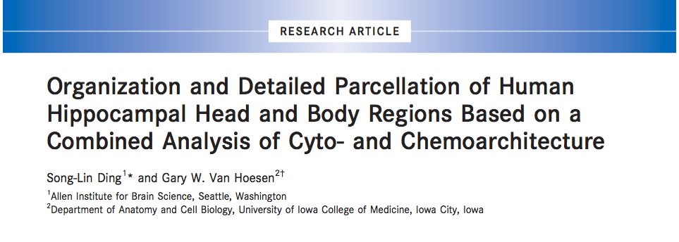
A newer ref worth mentioning here is:
Ding, & Van Hoesen. (2015). Organization and detailed parcellation of human hippocampal head and body regions based on a combined analysis of cyto‐and chemoarchitecture. Journal of Comp Neurol, 523(15), 2233-2253.
#SubfieldWednesday (7/n)
Ding, & Van Hoesen. (2015). Organization and detailed parcellation of human hippocampal head and body regions based on a combined analysis of cyto‐and chemoarchitecture. Journal of Comp Neurol, 523(15), 2233-2253.
#SubfieldWednesday (7/n)

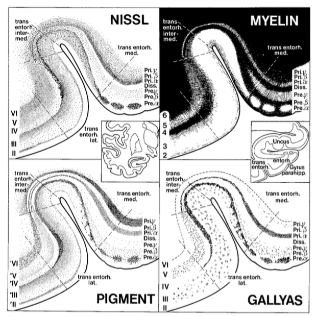
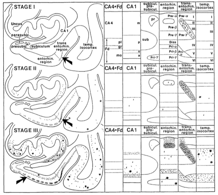
Ding & Van Hoesen provide unprecedented descriptions and visualizations of subfields in the hippocampal head-- including the mysterious uncal region!
#SubfieldWednesday (8/n)

#SubfieldWednesday (8/n)


Which neuroanatomy atlas do you use for your research?? Any others that we forgot?
Next week we will go over the most popular references for MTL cortex subregions.
#SubfieldWednesday (end)
Next week we will go over the most popular references for MTL cortex subregions.
#SubfieldWednesday (end)
unroll please, @threadreaderapp
• • •
Missing some Tweet in this thread? You can try to
force a refresh


