OK #VExUS #POCUS hemodynamicians, welcome back to the case. As promised, here are some follow up images:
@ArgaizR @ThinkingCC @khaycock2 @katiewiskar @Thind888 @MDBeni et al.
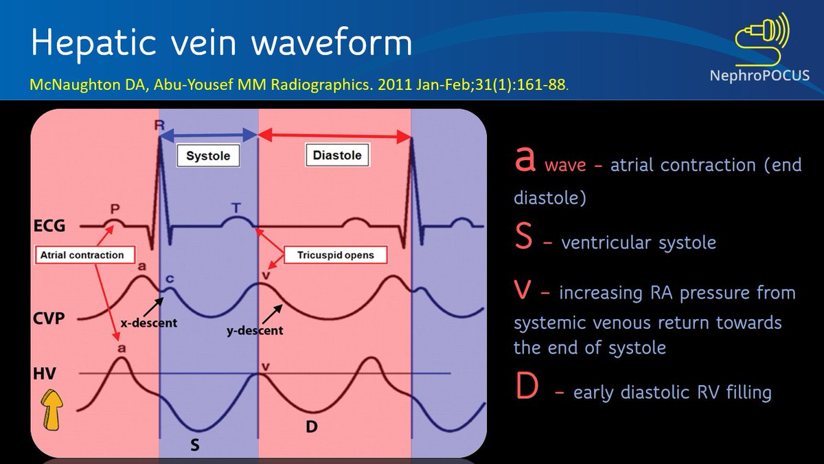
Hepatic v. πŸ‘‡
Looks D-only but could this be S-wave? (or a delayed D? Pt has Afib and predisposed to having smaller S but wondering if there is some S-D fusion here)
Forgot, here is the IVC. Similar to previous.
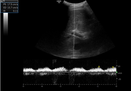
Portal vein #POCUS #VExUS
Interesting! Isn't it? (previously 100% pulsatile with flow interruptions)
Now the real twist:
* Patient did not get extra-UF for various reasons. I thought of repeating #POCUS anyways prior to discharge
* Why there seems to be improvement? Was the first scan 'too soon' after HD to appreciate flow changes? I thought flow changes should be quick πŸ€”
Don't know exact weight at the time of scan but at least 1 kg higher than of end-HD based on last available measurement (different scale).
Here is the PSAX you guys were asking for πŸ‘‡ Obtained in left lateral position.
M-mode. Kind of oblique but greyscale #POCUS seems to show septal flattening throughout the cycle. What u think @khaycock2 ?
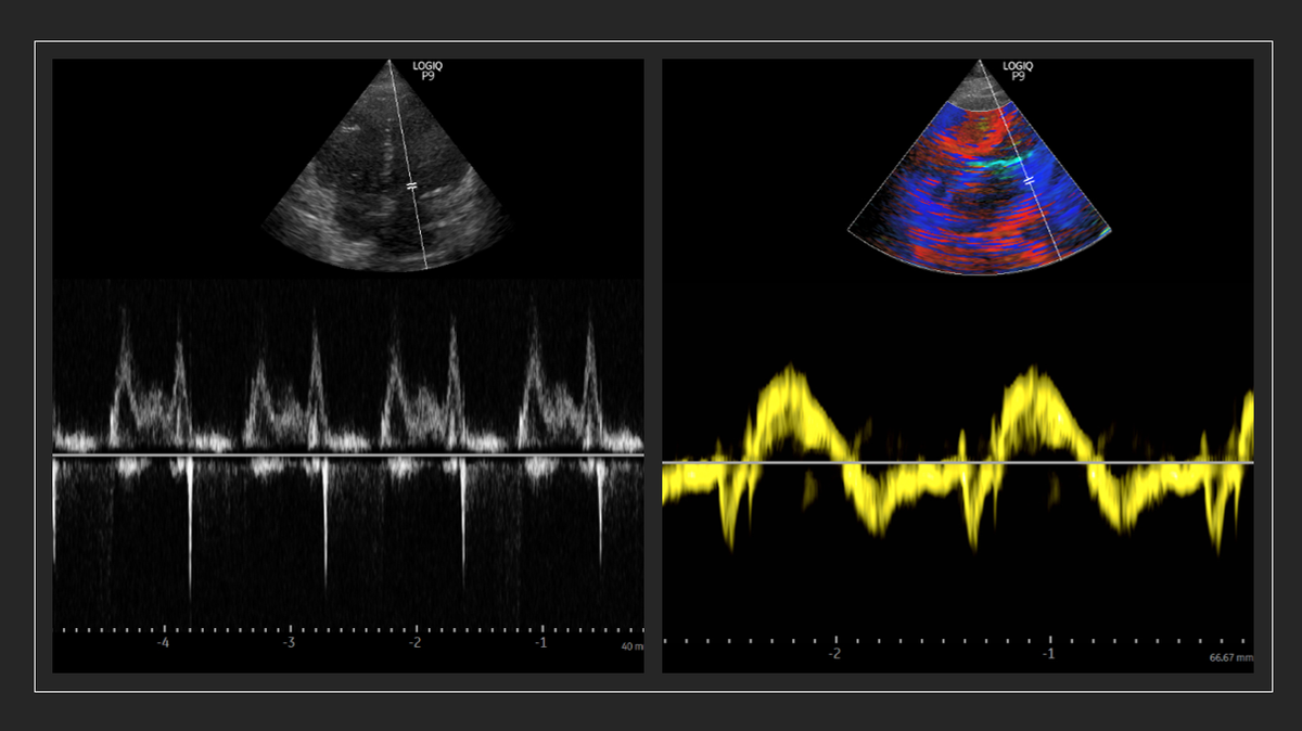
Subcostal SAX. No septal flattening? (different myocardial segments but does that alter D-sign?) #POCUS
Apical 4C. Likely more tilted angle of insonation making RV look bigger.
TR
TR jet and RVOT Doppler
Does this RVOT Doppler tell anything qualitatively? πŸ€”#POCUS
Septal TDI and Mitral inflow
@MDBeni, with respect to your question about management, cardiology will evaluate for TAVR as outpatient. They initiated work up including chest CT, which incidentally showed findings suggestive of chronic PE πŸ€”

β€’ β€’ β€’

Missing some Tweet in this thread? You can try to force a refresh
γ€€

Keep Current with NephroPOCUS

NephroPOCUS Profile picture

Stay in touch and get notified when new unrolls are available from this author!

Read all threads

This Thread may be Removed Anytime!

PDF

Twitter may remove this content at anytime! Save it as PDF for later use!

Try unrolling a thread yourself!

how to unroll video
  1. Follow @ThreadReaderApp to mention us!

  2. From a Twitter thread mention us with a keyword "unroll"
@threadreaderapp unroll

Practice here first or read more on our help page!

More from @NephroP

5 Feb
Renal #POCUS in a #dialysis patient: Looks fine at first glance (thin parenchyma as expected) but fanning anteriorly reveals a large cyst, which would have been missed otherwise. Labeled images and notes in thread πŸ‘‡
#Nephrology #MedEd #IMPOCUS
Labeled first image #POCUS
Note: peri-renal fat (usually prominent in ESRD patients) can be confused with free fluid.
Labeled second image #POCUS (obtained with phased array probe)
Note: EKG is not needed πŸ˜€
Read 5 tweets
11 Nov 20
#Nephrology consult for worsening creatinine in the context of diuresis in a patient with #heartfailure
Documented weight 3 kg ⬇️ than the previous day. No accurate urine output. Pt feels OK (has some unrelated issue).
Question: over diuresis?
#POCUS #MedEd #VExUS see thread πŸ‘‡
So, there is decreased LV systolic function and obviously diastolic dysfunction. Bi atrial enlargement. No significant pericardial effusion (there was trace on PLAX).
Next step in the hemodynamic circuit #POCUS assessment?
btw repeat weight was similar.
Next: IVC #POCUS assessment to assess right atrial pressure.
Here it is πŸ‘‡
Big and plethoric (take it as 15 mmHg in a spontaneously breathing person - we get RVSP of ~41 mmHg based on the above tricuspid gradient)
Read 10 tweets
25 Aug 20
#POCUS #IMPOCUS
Understanding #VExUS from scratch:
A mini #tweetorial on normal hepatic vein waveform genesis πŸ‘‡ #MedEd #echofirst

1/ Understanding color and direction
2/ What is S wave? #POCUS
3/ V wave #POCUS
Read 8 tweets
18 Aug 20
#POCUS Intra-renal vein Doppler waveform looking exactly like that of hepatic vein!
#VExUS obtained from a #nephrology clinic patient with pulmonary hypertension. #MedEd
Normal hepatic vein waveform #POCUS Image
Portal vein #POCUS in this patient - increased pulsatility. Image
Read 6 tweets
11 Jul 20
@Thind888 @FH_Verbrugge @khaycock2 Dear hemodynamic masters, saw a cirrhotic pt with oliguria (relatively preserved Scr) & pulmonary congestion. Based on these findings, suggested norepinephrine (pressors). Agree? any educational input appreciated.
Basically I interpreted as high output cardiac failure with elevated filling pressures leading to pulmonary congestion.
This tracing I believe is right intra-renal 'arterial' with diminished diastolic flow. Correct me if I'm wrong.
Sorry, didn't realize that the scale dropped
Read 4 tweets
18 Jun 20
1/ #Nephrology #POCUS case study:
Dr. X is rounding on an ESRD pt who initially presented with dyspnea after missing a dialysis (HD) session; underwent dialysis in the hospital. Pt asymptomatic at the time of exam and lung #ultrasound revealed πŸ‘‡ Further story in thread #MedEd
2/ Based on the 2-zone lung #POCUS, Dr. X orders for another session of HD. Notably, pt says he is at his 'dry weight' and HD nurse says they could only get 1.5L off during first session. Dr. X doesn't change his/her mind.
Info on various lung scan zonesπŸ‘‡
3/ Patient becomes hypotensive during HD and only ~500cc fluid could be removed.
Why can't we get more fluid out of a hypervolemic patient? Dr. X is perplexed and decides to more #POCUS Here is the IVC
Read 16 tweets

Did Thread Reader help you today?

Support us! We are indie developers!


This site is made by just two indie developers on a laptop doing marketing, support and development! Read more about the story.

Become a Premium Member ($3/month or $30/year) and get exclusive features!

Become Premium

Too expensive? Make a small donation by buying us coffee ($5) or help with server cost ($10)

Donate via Paypal Become our Patreon

Thank you for your support!

Follow Us on Twitter!