Today we continue our thread series discussing anatomical variability in the medial temporal lobe (MTL). Today’s topic is anatomical variability in the hippocampal head with a focus on the hippocampal digitations. 🍤🤓📢

#SubfieldWednesday (1/n)
But first, what do we mean by the hippocampal head? We are talking about the anterior part of the hippocampus that contains or is adjacent to the uncus.

#SubfieldWednesday (2/n) Figure 1 from https://doi.org/10.1038/nrn.2015.24 a| A schem
Now what about those “hippocampal digitations” ?

Ding & Van Hoesen (2015) describe external and internal digitations. The external digitations are the “bumps” that extend dorsally and the interior digitations are the “bumps” that extend ventrally.

#SubfieldWednesday (3/n) Figure 2A from https://doi.org/10.1002/cne.23786  HippocampaFigure 1B from https://doi.org/10.1016/j.neuroimage.2017.11.
According to Ding & Van Hoesen (2015), most hippocampi have 3 digitations, but some people have 2 or 4 (or more).

#SubfieldWednesday (4/n)
Q: Which subfields are contained in the digitations?
A: CA1 and subicular regions.

See figure below to see two examples of hippocampal subfields overlaid onto hippocampi with variable numbers of digitations.

#SubfieldWednesday (5/n) Fig. 5A and 5B. from https://doi.org/10.1016/j.neuroimage.20
Quick aside: It is important to remember that hippocampal digitations are different from “dentations” recently describes by Beattie et al, Neuropsychol, 2017.

doi.org/10.1016/j.neur…

We will have a different thread on those!

#SubfieldWednesday (6/n)
Now for the audience participation part! Have you come across any brains that have 4 or more hippocampal digitations? If so, comment below and post a screenshot!

#SubfieldWednesday (end)

• • •

Missing some Tweet in this thread? You can try to force a refresh
 

Keep Current with Hippocampal Subfield Group

Hippocampal Subfield Group Profile picture

Stay in touch and get notified when new unrolls are available from this author!

Read all threads

This Thread may be Removed Anytime!

PDF

Twitter may remove this content at anytime! Save it as PDF for later use!

Try unrolling a thread yourself!

how to unroll video
  1. Follow @ThreadReaderApp to mention us!

  2. From a Twitter thread mention us with a keyword "unroll"
@threadreaderapp unroll

Practice here first or read more on our help page!

More from @hipposubfields

14 Apr
Happy #SubfieldWednesday! Today we will be sharing a 🧵 on anatomical variability in the medial temporal lobe (MTL). MTL subregions vary in size and shape across individuals & these variations can make landmark identification and segmentation difficult

#SubfieldWednesday (1/n)
Today we will focus on the *most* consistent structure in the MTL: the hippocampal body. Most of our followers will be familiar with this canonical hippocampal body shape shown below in the coronal plane.

#SubfieldWednesday (2/n) Coronal plane from a T2 MRI depicting the canonical C shape
The shape is characterized by a consistent “C-shape” when viewed in the coronal plane. The C is formed by the cornu ammonus (CA) subfield on the lateral part of the hippocampal body.

#SubfieldWednesday (3/n)
Read 10 tweets
10 Feb
We are pleased to announce that today we have a guest #SubfieldWednesdsay 🧵 from @MarkCembrowski !

Check it out below!

(1/n)
When you look at a textbook diagram hippocampus, one sees a series of subfields - DG, CA3, and CA1. All of these regions have specialized properties relative to one another. But it raises the question: within each region, are the cell types uniform?

#SubfieldWednesdsay (2/n) Image courtesy of the Cembrowski lab: https://www.cembrowski
CA1 pyramidal cells of the rodent brain, one of the most studied neuron types in the brain, provide a good starting point to answer this question from both structural and functional perspectives.

#SubfieldWednesdsay (3/n) Image courtesy of Erik Bloss
Read 10 tweets
17 Dec 20
Is anyone planning to do some reading about hippocampal neuroanatomy over the holidays?

If you answered, "yes", this week's #SubfieldWednesday is for you! We will give you a list of "must read" atlas references about our favorite brain structure. 🍤❣️

#SubfieldWednesday (1/n)
In our 2015 paper (Yushkevich et al., NeuroImage, 2015), we provided a list of common atlases used for hippocampal subfield definition across labs.

#SubfieldWednesday (2/n) Table 2 from http://dx.doi.org/10.1016/j.neuroimage.2015.01.
One of the most commonly used atlases cited was:

Duvernoy, H. M. (2005). The human hippocampus: functional anatomy, vascularization and serial sections with MRI. Springer Science & Business Media.

#SubfieldWednesday (3/n)
Read 10 tweets
9 Dec 20
This week I thought I would share some terminology that you may or may not know!

#SubfieldWednesday (1/n)
Most of our followers probably already know how that the name "hippocampus" means "seahorse" in Greek.

#SubfieldWednesday (2/n) https://www.wbur.org/npr/505754756/the-seahorse-in-your-brai
What do you think? When it's dissected like this, I can definitely see the similarity!

#SubfieldWednesday (3/n) Hippocampus removed from the brain (left), in comparison to
Read 8 tweets
25 Nov 20
Hello and happy #SubfieldWednesday! Today we are going to get a bit more familiar with how the hippocampal subfields differ in their composition of different cell types, cell sizes, and layer thickness. 🍤🔬

#SubfieldWednesday (1/n)
Here are some images taken from five different hippocampal subfields (CA1, CA2, CA3, dentate gyrus, and subiculum). Can you tell which number corresponds to which subfield? 🤔

#SubfieldWednesday (2/n) Five different screenshots of histology slices of five diffe
Because a Nissl stain was applied to these slices, the cell bodies appear dark purple. This allows neuroanatomists to characterize the size, shape, and relative spacing of the cells.

#SubfieldWednesday (3/n)
Read 12 tweets
11 Nov 20
Happy #SubfieldWednesday! After a two-week hiatus we are returning to our quiz about the mysterious transentorhinal cortex!

This quiz even stumped some of us at @hipposubfields headquarters! We had to contact a neuroanatomist to confirm which answer is correct! (or at least "the most correct")
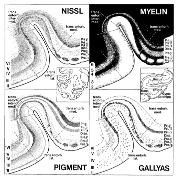
Braak and Braak (1985) originally described the transentorhinal cortex as a 'transition region between entorhinal cortex and temporal isocortex. This rules out answer A (part of ERC) Figure 3. from Braak H, Bra...
Read 11 tweets

Did Thread Reader help you today?

Support us! We are indie developers!


This site is made by just two indie developers on a laptop doing marketing, support and development! Read more about the story.

Become a Premium Member ($3/month or $30/year) and get exclusive features!

Become Premium

Too expensive? Make a small donation by buying us coffee ($5) or help with server cost ($10)

Donate via Paypal Become our Patreon

Thank you for your support!

Follow Us on Twitter!

:(