🧵In our paper Intraventricular Vector Flow Imaging with Blood Speckle Tracking in Adults: Feasibility, Normal Physiology and Mech… we use a new method, not only BST, and can be applied on adult probes. pubmed.ncbi.nlm.nih.gov/34620522/
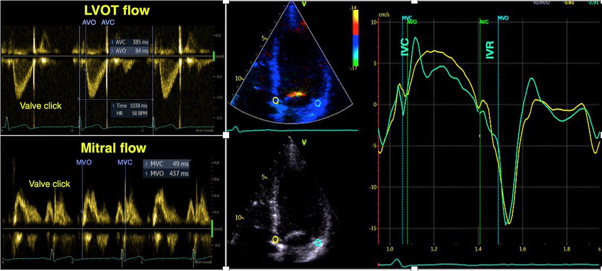
The main aim was to investigate the normal adult, intraventricular blood flow throughout the whole cardiac cycle, to compare with pw and colour Doppler M-mode and wall mechanics. (2D images courtesy of AS Daae).
As tweeted before, during IVR, there is simultaneous shortening of the base and elongation of the apex, inducing a volume shift with intraventricular apical flow, imparting a momentum and kinetic energy towards apex before start of early filling. This is thus *not* "wasted work"
1/ At the time of MVO and start of inflow, the AV plane also starts to move basally. This expands the space in the LVOT and behind the mitral ring, thus diverting inflowing blood laterally.
2/ This motion diverts blood flow downwards, creating vortices, counterclockwise in the LVOT, clockwise in the lateral part when viewed in a conventional 4-chamber view. The LVOT vortex is the largest as there is more room here.
3/ This diversion of blood flow, and start of diastolic vortex, is due to the ventricle being wider than the orifice, plus the expansion of the basal space, and thus starts near the LV base, as can be easily seen by CMM.
4/ The diverted flow into the LVOT is easily seen by pw Doppler, if you look for it, and is thus a normal phenomenon, although the E/A ratio in the LVOT will reflect the mitral E/A ratio, so a low E/A gives rise to the J-wave, which in reality is a high ALVOT.
5/ contrary to what has been published previously, this is not return flow from the apex. In our study, there was no delay between mitral E and e', while ELVOT had a delay of 116 ms. With an E of 82 cm/s, return flow from the apex would arrive after about 2 s.
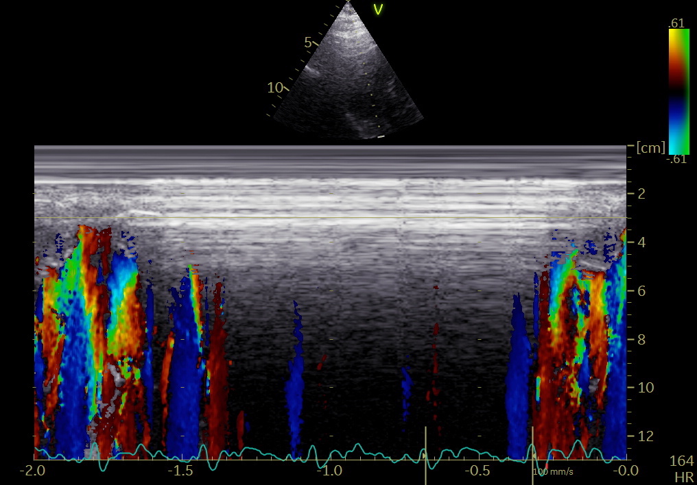
6/ during early filling, the lateral vortex being smaller and clockwise, seem to be extinguished, while the counterclockwise LVOT vortex expands (LVOT continuing to expand basally and more blood being diverted basally, as well as inflowing blood being diverted closer to the apex)
7/ This has previously been described as vortex propagation pubmed.ncbi.nlm.nih.gov/7867035/
but is actually an expansion, and apical propagation can be seen by colour M-mode; both negative vectors along the septum and positive vectors along the mid-lateral ventricle.

• • •

Missing some Tweet in this thread? You can try to force a refresh
 

Keep Current with Asbjørn Støylen

Asbjørn Støylen Profile picture

Stay in touch and get notified when new unrolls are available from this author!

Read all threads

This Thread may be Removed Anytime!

PDF

Twitter may remove this content at anytime! Save it as PDF for later use!

Try unrolling a thread yourself!

how to unroll video
  1. Follow @ThreadReaderApp to mention us!

  2. From a Twitter thread mention us with a keyword "unroll"
@threadreaderapp unroll

Practice here first or read more on our help page!

More from @strain_rate

12 Oct
🧵Having previously tweeted on the post ejection LV velocities and the relation to aortic valve closure mechanics,
from findings in our recent paper
onlinelibrary.wiley.com/doi/epdf/10.11…
I will now take a look on the isovolumic relaxation.
1/ In the paper pubmed.ncbi.nlm.nih.gov/34184410/ we found a consistent finding of isovolumic apical lengthening and basal shortening
2/ This, of course, indicates a volume short, from base to apex, even if total volume is constant.
Read 11 tweets
31 Aug
Apart from the physiological implications, what are the consequences of this study onlinelibrary.wiley.com/doi/epdf/10.11… for timing of valve openings and closures by tissue Doppler?
1/ Valve closures can be timed by tissue Doppler and mitral ring motion. However, only the septal motion will reliably show AVC.
2/ MVO is close to the END of the pre ejection spike. Timing MVC by the start of the pre ejection spike will result in an error of about 40 ms too early. Timing by the peak R wave will result in about the same error.
Read 13 tweets
26 Aug
1/ our paper elucidates the end ejection tissue velocity spike as well, and relation to AVC.
onlinelibrary.wiley.com/doi/epdf/10.11…
The negative velocity post ejection spike had an average duration of 35 ms, ending about 10 ms after AVC in the septum. Thus, this spike is not isovolumic relaxation, and the true IVR (AVC to MVO) is from the end of the spike to start of mitral flow.
The close relation between AVC and the end of the pre ejection spike has been shown before by phonocardiography pubmed.ncbi.nlm.nih.gov/16364793/ , and by high frame rate B-mode of the aortic valve pubmed.ncbi.nlm.nih.gov/20331696/ .
Read 12 tweets
20 Jul
1/ Our paper on pre- and post ejection tissue velocities by #EchoFirst in relation to valve closures and openings is out: onlinelibrary.wiley.com/doi/epdf/10.11…
2/ 22 healthy subjects, Valve openings and closures timed by Doppler flow, and transewfrred to Tissue Doppler recordings. Image
3/ Pre ejection velocities started 24.8 ms after start QRS, with a duration of 51.5 ms, ending about 11.5 ms after MVC. Thus, both electromechanical delay and pre ejection velocity occurs *before* onset of IVC, and are not a measure of IVC or Isovolumic acceleration. Image
Read 12 tweets
19 Mar
1/ Man ca 30, exertional dyspnea, CPET with normal VO2max, but pulmonologist concerned about possible drop in CO at peak exercise. Normal resting echo, no LVOT obstruction or gradient, no MR. Dobutamine stress: Chordal SAM, no regional ischemia
2/ Intraventricular gradient
3/ shown by CMM to be mid ventricular, moving towards apex in systole. No concomitant MR.
What next?
Read 7 tweets
27 Jan
1/ #LBBB generates often a classical pattern on #EchoFirst. The pattern is very distinctive in Tissue Doppler of the septum.
The classical pattern arises from the time lapse of the activation and relaxation of the two walls, creating a pattern of interaction due to a sequence temporal imbalances of the tension between the two walls.
2/ As the septum is activated first, it contracts (shortening - septal flash) without activation of the lateral wall, which stretches. This generates slower pressure build up than a normal IVC, which then is prolonged.
Read 10 tweets

Did Thread Reader help you today?

Support us! We are indie developers!


This site is made by just two indie developers on a laptop doing marketing, support and development! Read more about the story.

Become a Premium Member ($3/month or $30/year) and get exclusive features!

Become Premium

Too expensive? Make a small donation by buying us coffee ($5) or help with server cost ($10)

Donate via Paypal Become our Patreon

Thank you for your support!

Follow Us on Twitter!

:(