Humbling pleural procedure case to share.

70 y/o admitted with fever, hypoxia, R flank pain, loculated pleural effusion (right lower) on CXR. Concern for empyema prompting abx, chest US and possible intervention.

How would you manage? (poll to follow)

#POCUS #IMPOCUS

1/
2/
It is overnight on the ward. Which of the following would be your advised management?
3/
Seemed like a reasonable-sized pocket, and had signs of complexity (hyperechoic content, did not appear to be "free flowing). Given this and concern for empyema, plan was for thoracentesis, and if purulence or high risk features on fluid analysis, convert to pigtail.
4/
Thoracentesis was attempted using long 18 g angiocath, but no fluid was able to be aspirated. Live ultrasound guidance was used, which verified needle in the intended location.
(Image is undergained, turn up screen brightness to see)

5/
still image of needle in intended location
6/
What would be your next step?
7/
Thought was that fluid may have been too thick for aspiration w 18g. Attempted w 8Fr kit same site. Still no fluid aspirated. procedure was ended.

CT - thickened pleura, minimal pleural effusion, air locule at thora site. Later PET revealed FDG uptake in thickened pleura.
8/
Pt underwent VATS pleural biopsy which revealed mesothelioma. It turned out that fevers were due to extrathoracic source (R sided pyelonephritis), infection improved with abx.
9/
Some reflections
-Malignant pleural thickening can mimic pleural effusion on ultrasound
-Live US guidance can help verify needle position in challenging cases
-Can we distinguish pleural thickening from pleural effusion on ultrasound?

10/
There are reports of using color flow to distinguish pleural effusion from pleural thickening
sciencedirect.com/science/articl…
The study showed better specificity and PPV using CF vs gray scale. False negatives occurred due to color gain too low (or lack of movement in pleural fluid)
11/
And a review on pleural effusion vs thickening: touchrespiratory.com/airway-and-lun…
12/
Thoughts? Would you have done the thora?
In retrospect, procedure was not beneficial, and created potential risk. But it was difficult to know that at the time, and there was a compelling indication. Having encountered this scenario, can we discern such cases in the future?
13/

• • •

Missing some Tweet in this thread? You can try to force a refresh
 

Keep Current with IM POCUS Focus

IM POCUS Focus Profile picture

Stay in touch and get notified when new unrolls are available from this author!

Read all threads

This Thread may be Removed Anytime!

PDF

Twitter may remove this content at anytime! Save it as PDF for later use!

Try unrolling a thread yourself!

how to unroll video
  1. Follow @ThreadReaderApp to mention us!

  2. From a Twitter thread mention us with a keyword "unroll"
@threadreaderapp unroll

Practice here first or read more on our help page!

More from @IMPOCUSFocus

25 Dec 20
A tweetorial on optimal machine settings for B line image acquisition, with a focus on: focal position at pleura, harmonics off, and increased far gain.

Inspired/based on the recent article - onlinelibrary.wiley.com/doi/full/10.10…
@cameron_baston @IsaacMatthiasM1

#POCUS #IMPOCUS

1/18
Let's start with some polls
We will stick to low frequency probes here (i.e. curvilinear and phased array - linear has its own purpose for detailed pleural assessment)

Which preset do you prefer for B line assessment?

2/
What depth do you typically set?

3/
Read 19 tweets
22 Nov 20
1/
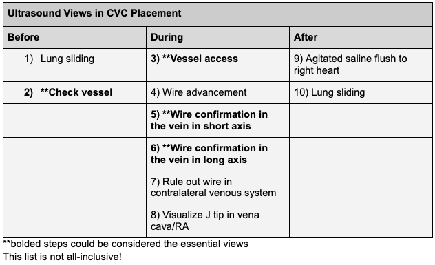
Thread: Ultrasound for CVC placement - more than just follow the needle tip and confirm the wire.

There are many potential uses of ultrasound during CVC placement, shown here. Not all are essential.

#POCUS #IMPOCUS #FOAMED #FOAMUS Image
2/
Pre-procedure
Lung sliding on ipsilateral side - for a comparison to post-procedure
-use linear probe
-apical and lateral/anterior
3/
Check vessel
-confirm compressibility
-understand axis of vessel
-check prox and distal for stenoses
-optimize depth and gain (this image may be undergained)
Read 25 tweets
15 Nov 20
Let's talk about placement of small bore nasoenteric feeding tubes and use of ultrasound for this (A thread)

Scenario: Pt with gastroparesis and COVID/ARDS, planning to prone, want post-pyloric tube. No dedicated team for this. Can we place safely w #POCUS guidance?

1/
How is small bore feeding tube (SBFT) different from standard NG?

Narrower (6-8 Fr vs 14-18 Fr), more flexible (but w rigid stylet for placement), longer. Thus: more comfortable, gastric or post-pyloric, ineffective for suction, prone to clogging, prone to PTX if placed in lung.
Some methods used for placement
1) Blind placement - NOT RECOMMENDED. 1-2% risk of PTX
2) 2 radiograph method (safe but time consuming)
3) Capnometry
4) Endoscopic visualization
5) Electromagnetic
6) Fluoroscopic
7) Ultrasound?

Nice article on this: med.virginia.edu/ginutrition/wp…

3/
Read 31 tweets
9 Mar 20
1/
Happy Monday everyone! We have a special case for you today written by UPMC PGY2 #POCUS enthusiast @MikeTao15.

A 52 yo M with hx of EtOH/HCV cirrhosis, VTE on Eliquis, HFrEF presents w worsening abd distention and pain. He is disoriented and unable to give much history.
2/
Cirrhosis previously complicated by esophageal varices, hepatic encephalopathy, and ascites. No follow up since last hospital admission at outside facility ~1 year ago. Has not been taking meds.

Vitals in the ED: Temp 38.1, HR 90, BP 98/70, Pulse Ox 96% on 2L
3/
He is lethargic, arousable to voice and oriented to self only
+scleral icterus
Cardiac + pulmonary exams normal
Abdomen distended, diffusely tender, no rebound/guarding
2+ b/l edema to thighs
Jaundiced, scattered ecchymoses
+asterixis
Read 36 tweets
28 Feb 20
Ever found DVT POCUS challenging? We do.

Here is a #Tweetorial on #POCUS for Lower Extremity DVT assessment. Let’s walk through a case and discuss common questions that arise.

50s f no PMH presenting to the ED on a Friday evening with right leg swelling and redness...

1/
History/exam/labs:

No travel, surgeries, immobilization. No dyspnea, CP. No meds. No FH of VTE. no tobacco.

HR 88 SpO2 98% RA BP 160/90 T 37.

Exam unremarkable other than 2+ LE edema erythema and tenderness from right ankle to knee.

CBC/BMP/coags normal

2/
Wells-DVT score 1 (for pitting edema confined to symptomatic leg), with a moderate pre-test probability for DVT.

It is Saturday. Formal vascular study (FVS) is not available until Monday. D-dimer is considered. Would you check D-dimer in this case? (poll)

3/
Read 39 tweets
11 Jan 20
1/
After a break for the new year, we are back with a case of #POCUS and abdominal distension.

59 y/o m with no medical history presenting with increasing abdominal girth and now altered mental status.

#IMPOCUS
2/
HPI:
well until 3 months ago. Since then, progressive fatigue, nausea, mild diffuse itching without rash. Few weeks of early satiety, poor PO intake, and now increasing abdominal girth. Last BM 3 days ago. No vomiting. Weight unchanged over 3 months. No fevers/chills.
3/
No home meds. 2-3 etoh drinks per day (none for a few wks). Unable to work due to symptoms. Past 1-2 days he has become confused and unable to ambulate.
Read 30 tweets

Did Thread Reader help you today?

Support us! We are indie developers!


This site is made by just two indie developers on a laptop doing marketing, support and development! Read more about the story.

Become a Premium Member ($3/month or $30/year) and get exclusive features!

Become Premium

Too expensive? Make a small donation by buying us coffee ($5) or help with server cost ($10)

Donate via Paypal

Or Donate anonymously using crypto!

Ethereum

0xfe58350B80634f60Fa6Dc149a72b4DFbc17D341E copy

Bitcoin

3ATGMxNzCUFzxpMCHL5sWSt4DVtS8UqXpi copy

Thank you for your support!

Follow Us on Twitter!

:(