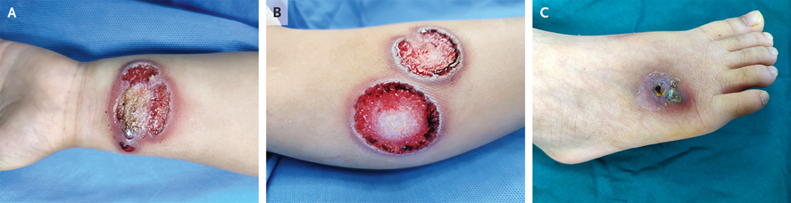
A 12-MO♀️ girl, immunization for hepatitis B, 2 weeks prior: a rapidly spreading, nonblanching, nonpalpable, purpuric rash over legs & cheeks surrounded by edema, 37.6°C, nontoxic in appearance

Leukocytosis,⬆️CRP
1/9

#dermatology #Emergency #Pediatrics doi.org/10.1093/cid/ci…
The next day, the hemorrhagic rash became palpable
Pharyngeal PCR:➕for enterovirus
🔬 of purpuric lesions: leukocytoclastic vasculitis, & DI studies showed vascular wall fibrinogen deposition, consistent with ACUTE HEMORRHAGIC EDEMA OF INFANCY (AHEI)
2/9

#pediatric #IDtwitter
Typical patient of ACUTE HEMORRHAGIC EDEMA OF INFANCY:
✔️6–24 months of age,
✔️during winter,
✔️nontoxic presentation,
✔️low-grade fever,
✔️abrupt onset of large purpuric skin lesions,
✔️and edema in face and extremities
3/9

doi.org/10.1093/cid/ci…
#MedTwitter #pediatria
ACUTE HEMORRHAGIC EDEMA OF INFANCY is a self-limited disease and the prognosis is excellent.

Complete recovery usually occurs within 1–3 weeks
4/9

#MedStudentTwitter #MedicalStudents
ACUTE HEMORRHAGIC EDEMA OF INFANCY
✔️associated with some viruses & bacteria (adenovirus, cytomegalovirus, herpes simplex virus, varicella zoster virus, tuberculosis, streptococci, staphylococci, pneumococcus)
✔️but the etiology of this disease remains unknown
5/9

#microbiology
AHEI triggered by:
📌infection (respiratory infection, urinary tract infection, etc.), vaccination (H1N1, MMR, BCG, DTaP, polio, H. influenzae), or
📌drug intake (penicillin, cephalosporin, TMP/SMX, etc.),
which reveal a possible immune-mediated pathophysiology
6/9

#Doctor
The girl had enterovirus isolated from the throat, and hepatitis B vaccine was given 2 weeks before admission, signifying a possible association with acute hemorrhagic edema of infancy.
7/9

#microbiology
Diagnosis is based on clinical features but if diagnosis is unclear, a skin biopsy of the rash will be helpful.

The most common histopathological description is perivascular neutrophilic infiltration with nuclear fragments in the vascular wall and fibrinoid necrosis
8/9
Differential diagnosis includes Sweet syndrome, erythema multiforme, purpura fulminans, Kawasaki disease, & meningococcal septicemia

Acute hemorrhagic edema of infancy is a benign, self-limited leukocytoclastic #vasculitis of small vessels affecting #children <2 years of age
9/9

• • •

Missing some Tweet in this thread? You can try to force a refresh
 

Keep Current with Antonio Guerrero

Antonio Guerrero Profile picture

Stay in touch and get notified when new unrolls are available from this author!

Read all threads

This Thread may be Removed Anytime!

PDF

Twitter may remove this content at anytime! Save it as PDF for later use!

Try unrolling a thread yourself!

how to unroll video
  1. Follow @ThreadReaderApp to mention us!

  2. From a Twitter thread mention us with a keyword "unroll"
@threadreaderapp unroll

Practice here first or read more on our help page!

More from @guerreroespejo

Dec 15
Lactante♀️, 20 meses: fiebre, sialorrea, lesión costrosa hemática con contenido purulento y restos de costras melicéricas, eritema, edema y lesiones vesiculosas en 2º dedo de la mano, adenopatías epitrocleares, vesículas en paladar y aftas en encías
1/5

#pediatria #infecciosas Image
El PANADIZO HERPÉTICO es una lesión cutánea producida, especialmente en pediatría, por el virus herpes simple tipo 1.
2/5

pap.es/articulo/13681…
#AtencionPrimaria #microbiología
La presentación clínica habitual es una lesión con vesículas arracimadas, cuya localización típica es en primer o segundo dedo.

En los pacientes pediátricos es frecuente que se produzca tras autoinoculación desde una gingivoestomatitis herpética.
3/5

#MIR #EducaciónMédica
Read 5 tweets
Dec 12
A 65-YO man, from North African: asymptomatic dorsolumbar mass for 30 years with normal skin appearance.

MRI: ?
1/6

doi.org/10.1093/cid/ci…
#radiology #MedTwitter #parasitology
MRI: a multivesicular cyst in the latissimus dorsi muscle (arrow)

🧪for Echinococcus granulosus: ELISA, & Western blot➕
Surgery: multiple daughter vesicles of E. granulosus, with the presence of multiple scolices

INTRAMUSCULAR HYDATID CYST
2/6

#parasites #medicine #radres
No puncture should be performed, to avoid dissemination of the cysts that can cause an anaphylactic shock.

The patient received 3 other courses of albendazole: serologic control became negative after 1 year.
3/6

#IDtwitter #surgery
Read 6 tweets
Dec 10
A 35-YO♂️: a 9-month of white concretions encircling the hairs in both axillae (A).

A Wood’s lamp🔎: yellow-green fluorescence of the concretions (B).

Dermoscopy: cottonlike structures on the hair shafts (C).
1/6

DOI: 10.1056/NEJMicm2206453
#dermatology #dermatología Image
TRICHOMYCOSIS AXILARIS

✔️ a superficial bacterial infection of the hair of the axillae
✔️misnomer, given that it is caused by corynebacterium species as opposed to fungi.
2/6

#dermtwitter #microbiology
After 1 week of treatment with topical clindamycin and a daily benzoyl peroxide wash, the patient’s symptoms abated and did not recur.
3/6

#MedTwitter #MedStudentTwitter
Read 6 tweets
Dec 8
Lactante, 9 meses: lesiones peribucales, edema palpebral, conjuntivitis, xerosis peribucal con costras melicéricas, eritrodermia generalizada y lesiones en cuello, extremidades superiores y glúteos, con signo de Nikolsky +.
1/5

#pediatria #AtencionPrimaria #urgencias
SINDROME DE PIEL ESCALDADA ESTAFILOCÓCICA (Enfermedad de Ritter).

📌frecuente en < 5 años,
📌eritrodermia con signo de Nikolsky
📌conjuntivitis y edema facial con descamación periorificial.
2/5

continuum.aeped.es/screens/play/1…
@ContinuumAEP @aepediatria #infecciosas #microbología
@ContinuumAEP @aepediatria La enfermedad de Ritter está causada por cepas de estafilococo productoras de toxinas que se dirigen al complejo desmogleína 1 en la zona granulosa de la epidermis, provocando la exfoliación de la piel.
3/5

#MIR #MedTwitter
Read 5 tweets
Dec 5
A 59-YO DM, steroid, myasthenia gravis with thymoma: headache & deviation of tongue to the R side with fasciculation suggestive of right 12th cranial nerve palsy

CT: sphenoid sclerotic changes, soft tissue density lesion with calcification.
1/9

#neurology #radiology #IDtwitter Image
MRI brain: a hypointense lesion involving the body of sphenoid (R > L) and clivus on T2 weighted images and T1 weighted images with moderate contrast enhancement possibly fungal in aetiology.
2/9

doi.org/10.1016/j.idcr…
#radiologist #radres #MedTwitter ImageImage
Endoscopic sphenoidotomy & debridement: invasive fungal sphenoid sinusitis & CENTRAL SKULL BASE OSTEOMYELITIS involving the clivus

🔬from sphenoid mucosa: morphologically ASPERGILLUS with foci of tissue invasion.
3/9

#microbiology #maxillofacial #Orthopedics
Read 8 tweets
Dec 3
A 5-YO♂️ with a history of IgA vasculitis & a pustular nasal lesions had started scabbing after 2 days of systemic glucocorticoids: progressively painful skin ulcerations of L forearm (A), R calf (B) & R foot (C).
1/4

DOI: 10.1056/NEJMicm2204111
#Pediatrics #dermatology
Biopsy samples from all three lesions showed a dense neutrophilic infiltrate with negative tissue cultures.

PYODERMA GANGRENOSUM
2/4

#MedTwitter #Doctor #MedStudentTwitter
Laboratory & radiographic: no signs of underlying systemic disease, such as cancer, inflammatory bowel disease, or autoimmune conditions.

After 8 months of glucocorticoids: the lesions resolved completely.
¾

#resident #pediatric #MedicalStudents
Read 4 tweets

Did Thread Reader help you today?

Support us! We are indie developers!


This site is made by just two indie developers on a laptop doing marketing, support and development! Read more about the story.

Become a Premium Member ($3/month or $30/year) and get exclusive features!

Become Premium

Don't want to be a Premium member but still want to support us?

Make a small donation by buying us coffee ($5) or help with server cost ($10)

Donate via Paypal

Or Donate anonymously using crypto!

Ethereum

0xfe58350B80634f60Fa6Dc149a72b4DFbc17D341E copy

Bitcoin

3ATGMxNzCUFzxpMCHL5sWSt4DVtS8UqXpi copy

Thank you for your support!

Follow Us on Twitter!

:(