Daniel Gewolb, MD Profile picture
May 22 4 tweets 3 min read Twitter logo Read on Twitter
Case of emphysematous epiglottitis in an adult

Epiglottitis is an emergency as it can potentially cause airway compromise especially in children who have smaller airways #neurotwitter #ent #peds #Neurosurgery #MedTwitter #MedEd @ASHNRSociety ImageImage
▶️In children the diagnosis can be confirmed with upright plain film. CT requires placing the patient supine which may exacerbate inspiratory strider

▶️In adults, the diagnosis may not be suspected clinically so patients may end up with a CT scan as in this case
▶️Bacterial infection typically 2/2 H. Influenza in unvaccinated children

▶️In adults, possible pathogens include Strep, Staph, and H. influ
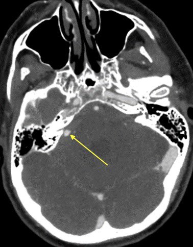
▶️CT shows emphysematous swelling of the epiglottis and marked thickening of the aryepiglottic folds (arrows 2nd image). The aryepiglottic folds delineate the anteromedial borders of the pyriform sinus ImageImage

• • •

Missing some Tweet in this thread? You can try to force a refresh
 

Keep Current with Daniel Gewolb, MD

Daniel Gewolb, MD Profile picture

Stay in touch and get notified when new unrolls are available from this author!

Read all threads

This Thread may be Removed Anytime!

PDF

Twitter may remove this content at anytime! Save it as PDF for later use!

Try unrolling a thread yourself!

how to unroll video
  1. Follow @ThreadReaderApp to mention us!

  2. From a Twitter thread mention us with a keyword "unroll"
@threadreaderapp unroll

Practice here first or read more on our help page!

More from @daniel_gewolb

May 23
Interesting case in this patient with acute right-sided weakness

#neurorad #neurotwitter #meded #Neurosurgery #Neurology @TheASNR @RSNA #medtwitter ImageImageImageImage
Can you determine the diagnosis off the CT?
▶️Initial non-con CT shows a 3cm hyperdense lobulated extra-axial mass in the expected region of the left MCA bifurcation, consistent with a giant aneurysm. There are associated peripheral calcifications

▶️ What is the cause of the surrounding hypodensity?
Read 10 tweets
May 21
Case of diffuse CSF seeding of tumor in this patient w/ WHO grade 4 diffuse hemispheric glioma

#NeuroTwitter #neurosurgery #Neurology #peds #futureradres @The_ASPNR #MedEd ImageImageImageImage
▶️Prospectively this mass was thought to be an embryonal tumor w/ multilayered rosettes given the marked diffusion restriction, hemorrhage, and lack of surrounding edema 🧠
Imaging:

▶️T2 shows a heterogenous slightly hyperintense mass w/ areas of hypointensity

▶️No surrounding edema/infiltrating tumor is seen on T2/FLAIR

▶️Fluid-fluid level is seen suggestive of hemorrhage (arrow) Image
Read 6 tweets
May 21
Learning case in this 40 y/o F with history of whole brain radiation as a child for brain tumor treatment
#NeuroRad #neurosurgery #Neurology @TheASNR #NeuroTwitter #meded #radres ImageImageImageImage
Brain radiation is a risk factor for the development of meningiomas …this patient subsequently developed multiple meningiomas including this large frontal meningioma Image
The mass was treated and immediate post op CT shows expected post op change with the resection cavity, some hemorrhage, and pneumocephalus Image
Read 9 tweets
May 19
Interesting case of complicated acute bacterial rhinosinusitis in this child with no PMH presenting w/ HA, fever & L sided weakness

#NeuroTwitter #ent #radres #neurosurgery @TheASNR @ASHNRSociety @PhilipRChapman1 #radres #futureradres ImageImageImageImage
CT shows opacification of the frontal and anterior ethmoid sinuses without evidence of cortical dehiscence

💡 In peds, infection can spread through vascular channels w/o destroying the bone ImageImage
MR shows abnormal signal filling the sinuses with associated restricted diffusion 2/2 purulent material

Post contrast we see areas of relative hypoenhancement in comparison to the normal mucosa on the contralateral side ImageImageImage
Read 9 tweets
Mar 27
What is the most likely diagnosis in this adolescent with seizure? 🧠

(Sorry I have no CT without)

#neurotwitter #peds #Neurosurgery #Neurology @The_ASPNR @TheASNR #MedTwitter
What is the most likely diagnosis?
Answer: Confirmed supratentorial ependymoma

Predicting tumors is incredibly challenging in the absence of specific features …some learning points on the case in 🧵
Read 8 tweets
Mar 27
Case of a radiation induced pseudoaneurysm in this patient with headache and AMS 🧠

Imaging in thread #Neurosurgery #Neurology #neurotwitter #radres #MedEd #MedTwitter @TheASNR ImageImageImageImage
▶️Initial head CT shows subarachnoid hemorrhage centered in the right cerebellopontine angle cistern

▶️CTA confirms an aneurysm of the right anterior inferior cerebellar artery (AICA) ImageImage
▶️MR displays and ice cream shaped enhancing mass extending through the right internal auditory canal into the cerebellopontine angle cistern, consistent with a vestibular schwannoma #icecream

▶️Careful search into the history confirms the schwannoma was treated with radiation ImageImage
Read 4 tweets

Did Thread Reader help you today?

Support us! We are indie developers!


This site is made by just two indie developers on a laptop doing marketing, support and development! Read more about the story.

Become a Premium Member ($3/month or $30/year) and get exclusive features!

Become Premium

Don't want to be a Premium member but still want to support us?

Make a small donation by buying us coffee ($5) or help with server cost ($10)

Donate via Paypal

Or Donate anonymously using crypto!

Ethereum

0xfe58350B80634f60Fa6Dc149a72b4DFbc17D341E copy

Bitcoin

3ATGMxNzCUFzxpMCHL5sWSt4DVtS8UqXpi copy

Thank you for your support!

Follow Us on Twitter!

:(