1/ Fascinating new🧠#COVID study published in @ScienceAdvances
Haven't done a 🧵 like this in a while, but let’s break down what it does and doesn’t say
#SARSCoV2 infection & viral fusogens cause neuronal & glial fusion that compromises neuronal activity science.org/doi/10.1126/sc…
Haven't done a 🧵 like this in a while, but let’s break down what it does and doesn’t say
#SARSCoV2 infection & viral fusogens cause neuronal & glial fusion that compromises neuronal activity science.org/doi/10.1126/sc…
https://twitter.com/WesElyMD/status/1668214866551742467

2/ The authors pose the question regarding potential neuropathological mechanisms other than neuronal cell death that help viruses spread infection within the host that then leads to brain dysfunction
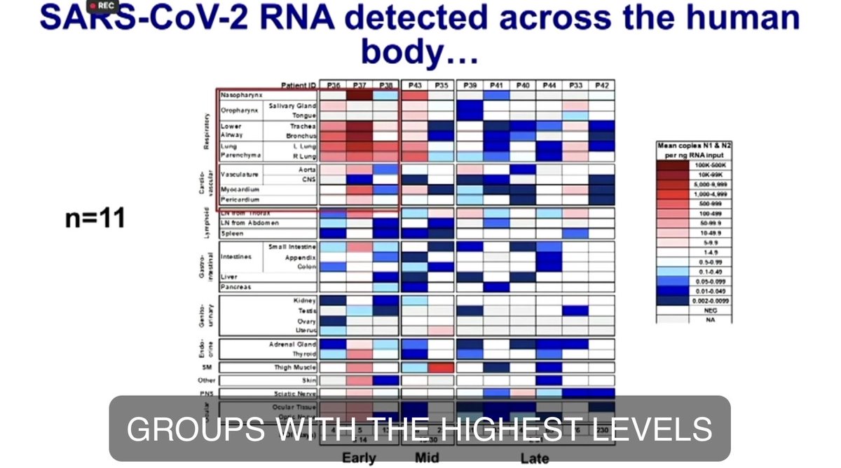
⚠️Brain damage can be caused without infecting neurons
doi.org/10.1002/jmv.26…
⚠️Brain damage can be caused without infecting neurons
doi.org/10.1002/jmv.26…
3/ What they found is VERY intriguing.
Instead of multiplying inside the cell and requiring the cell to burst & die in order to spread virions, the virus actually keeps the cell alive & uses it like a little trojan horse that docks on other similar neurons & fuses with them to
Instead of multiplying inside the cell and requiring the cell to burst & die in order to spread virions, the virus actually keeps the cell alive & uses it like a little trojan horse that docks on other similar neurons & fuses with them to
4/ then spread & replicate.
The virus doesn't have to leave the cell again & go into the extracellular space to reinfect another cell.
Because the virus stays within the cell & fuses with another, it can avoid detection by the immune system (hijacking the cell like a zombie)
The virus doesn't have to leave the cell again & go into the extracellular space to reinfect another cell.
Because the virus stays within the cell & fuses with another, it can avoid detection by the immune system (hijacking the cell like a zombie)
5/ Good for the virus, not so good for the host.
Here’s the catch. That cell is a zombie. It doesn’t work like it used to. And when it fuses with other cells those cells now don’t work well either.
The carefully crafted circuitry and subsequent cognitive function is disrupted.
Here’s the catch. That cell is a zombie. It doesn’t work like it used to. And when it fuses with other cells those cells now don’t work well either.
The carefully crafted circuitry and subsequent cognitive function is disrupted.
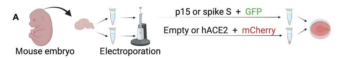
6/ FIRST UP: CELL CULTURE (MODIFIED MOUSE 🐭🧠)
- Dissected mouse brains (hippocampus) that were modified to express human ACE2
- Zapped the cells w/ electricity⚡️to to make them more permeable to add a plasmid that binds to hACE2 & marked w/ green fluorescent protein (GFP)
- Dissected mouse brains (hippocampus) that were modified to express human ACE2
- Zapped the cells w/ electricity⚡️to to make them more permeable to add a plasmid that binds to hACE2 & marked w/ green fluorescent protein (GFP)
7/ - Repeated the same thing with a 2nd population of hippocampal cells but instead of fluorescent green they made these ones red (mCherry🍒).
- Added color to the cells so they could see direct transfer of cellular material if fusion did occur.
- Added color to the cells so they could see direct transfer of cellular material if fusion did occur.
8/ - Put both green & red modified mouse brain cells on the same plates 🧫
- Grew the cultures in the lab x 5 days 👩🔬
- Then added virus or placebo to the cultures and let them “cook” x 72 hours 🍳
- Fixed and examined cultures under the microscope 🔬
- Grew the cultures in the lab x 5 days 👩🔬
- Then added virus or placebo to the cultures and let them “cook” x 72 hours 🍳
- Fixed and examined cultures under the microscope 🔬

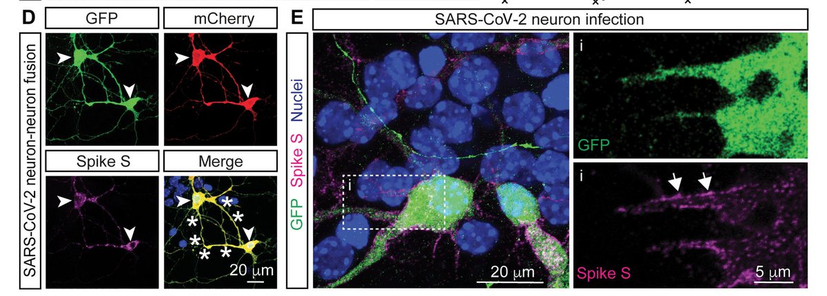
9/ Under the microscope they saw fusion of infected neurons (characterized by the presence of both the GFP (green) and mCherry (red) fluorescent proteins appearing in both neurons (yellow in merge) for all the SARS-CoV-2-titers used.

10/ They also stained the cells for Spike (S) protein, which was present on the surface of infected fused neurons.
No neuronal fusion or presence of Spike protein was seen in the control cultures
Glial cells expressing hACE2 were also positive for the spike protein staining.
No neuronal fusion or presence of Spike protein was seen in the control cultures
Glial cells expressing hACE2 were also positive for the spike protein staining.
11/ In addition to neuron-neuron, they also observed other fusion phenotypes, including neuron-glia and glia-glia.
High doses of neuronal SARS-CoV-2 infection resulted in cell damage, which was not apparent at the lowest doses.
⚠️Infectious DOSE matters (but we knew that😷)
High doses of neuronal SARS-CoV-2 infection resulted in cell damage, which was not apparent at the lowest doses.
⚠️Infectious DOSE matters (but we knew that😷)
12/ NEXT UP: BRAIN ORGANOIDS (HUMAN)
Used 43- to 50-day-old “mini brains” or human embryonic stem cell (hESC)–derived 3D brain organoids
Several other labs have used 🧠organoids h/t @VirusesImmunity
Neuroinvasion of SARS-CoV-2 in human and mouse brain
doi.org/10.1084/jem.20…
Used 43- to 50-day-old “mini brains” or human embryonic stem cell (hESC)–derived 3D brain organoids
Several other labs have used 🧠organoids h/t @VirusesImmunity
Neuroinvasion of SARS-CoV-2 in human and mouse brain
doi.org/10.1084/jem.20…
13/ The other paper that has stuck with me was the one from December 2020 about the choroid plexus organoids.
SARS-CoV-2 Infects the Brain Choroid Plexus and Disrupts the Blood-CSF Barrier in Human Brain Organoids h/t @Mad_Lancaster
doi.org/10.1016/j.stem…
SARS-CoV-2 Infects the Brain Choroid Plexus and Disrupts the Blood-CSF Barrier in Human Brain Organoids h/t @Mad_Lancaster
doi.org/10.1016/j.stem…
14/ Organoids were then transduced with adeno-associated virus (AAV) expressing GFP (green marker) and “cooked” x 10 more days
Same as prior experiment, they then infected them with #SARSCoV2 and cooked x 72 hours
Then fixed and looked under microscope 🔬
Same as prior experiment, they then infected them with #SARSCoV2 and cooked x 72 hours
Then fixed and looked under microscope 🔬

*Lunch Break*
Have you eaten yet today?
Here's a gentle reminder to get some food, hydrate, and stretch a bit 😊
Have you eaten yet today?
Here's a gentle reminder to get some food, hydrate, and stretch a bit 😊
15/ Longer break than expected, now BACK to this awesome study!
Similar to what they observed after infection of 2D neuronal mouse brain cultures, they found that SARS-CoV-2–infected human brain organoids exhibited neuronal syncytia formed by GFP-interconnected neurons
Repeat📸
Similar to what they observed after infection of 2D neuronal mouse brain cultures, they found that SARS-CoV-2–infected human brain organoids exhibited neuronal syncytia formed by GFP-interconnected neurons
Repeat📸

16/ Syncytia are evolutionarily conserved cellular structures that form by the multiple cell fusions of cells with single nuclei.
Here's a study in @Nature looking at synctia formation during #SARCOV2 in the lung and relationship to lymphocytes:
nature.com/articles/s4141….
Here's a study in @Nature looking at synctia formation during #SARCOV2 in the lung and relationship to lymphocytes:
nature.com/articles/s4141….
17/ Importantly, cell-to-cell contact and transfer has already been demonstrated in other viruses like human immunodeficiency virus (HIV) (type 1 and 2) and human T-cell lymphotropic virus type 1 (HTLV-1) as a mechanism to spread infection. h/t @dbdugger
ncbi.nlm.nih.gov/pmc/articles/P…
ncbi.nlm.nih.gov/pmc/articles/P…
18/ So then they were like, “whoa OK, but can we cause this same neuronal cell fusion by just exposing host cells to isolated fusogen protein and not the whole virus?”
So they then used a known fusogen
👉p15 from baboon orthoreovirus (BRV)
AND
👉 Spike S protein from #SARSCoV2
So they then used a known fusogen
👉p15 from baboon orthoreovirus (BRV)
AND
👉 Spike S protein from #SARSCoV2
19/ ⚠️IMPORTANT NOTE:
BRV infects brain of primates causing meningoencephalomyelitis.
BUT unlike spike S protein, p15 is the only viral protein required by BRV to form a syncytium, with no receptor protein on the host cell being needed to mediate fusion.
pubmed.ncbi.nlm.nih.gov/33443166/
BRV infects brain of primates causing meningoencephalomyelitis.
BUT unlike spike S protein, p15 is the only viral protein required by BRV to form a syncytium, with no receptor protein on the host cell being needed to mediate fusion.
pubmed.ncbi.nlm.nih.gov/33443166/
20/ SO, they isolated the mouse brain cells again but this time zapped ⚡️them with a plasmid containing the p15 protein & GFP (green). And then repeated the same process using mCherry (red) with a control vector.
Plated them together again and let cultures cook x 7 days. 🧫
Plated them together again and let cultures cook x 7 days. 🧫
21/ They revealed p15 was sufficient to induce neuronal fusion by again seeing the presence of neurons containing both the GFP and mCherry fluorescent proteins (yellow in merge).
Notably again, this phenotype was NEVER observed with the control vector (without p15)!
Notably again, this phenotype was NEVER observed with the control vector (without p15)!

22/ To further see if the fusion and diffusion between cells was due to the fusogenic properties of p15, they created an inactive mutant version by editing the transmembrane domain.
They repeated the experiment with the inactive version of p15 & no neuronal fusion was observed!
They repeated the experiment with the inactive version of p15 & no neuronal fusion was observed!

23/ AGAIN, unlike p15, Spike protein must bind to the hACE2 receptor to trigger fusion (requires BOTH spike S & hACE2 to be expressed).
Did a similar experiment & zapped the humanized mouse brain cells with a plasmid containing a codon-optimized version of Spike protein + GFP.
Did a similar experiment & zapped the humanized mouse brain cells with a plasmid containing a codon-optimized version of Spike protein + GFP.

24/ - They repeated this with another plasmid containing the hACE2 receptor and expressing mCherry (red).
- Plated them together and cultured x 7 days 🧫
- Expression of both Spike and hACE2 resulted in the fusion of adjacent neurons and the mixing of the fluorescent proteins🔬
- Plated them together and cultured x 7 days 🧫
- Expression of both Spike and hACE2 resulted in the fusion of adjacent neurons and the mixing of the fluorescent proteins🔬

25/ The expression of either Spike protein *or* hACE2 alone did not generate any fusion events.
⚠️The presence of *BOTH* the fusogen and its specific receptor (hACE2) was *required* to initiate cellular fusion (See First Row)
Repeated 📸 of figure in last 3 tweets
⚠️The presence of *BOTH* the fusogen and its specific receptor (hACE2) was *required* to initiate cellular fusion (See First Row)
Repeated 📸 of figure in last 3 tweets

26/ Just like with the P15, they wanted to test if the fusion and diffusion between cells was due to the fusogenic properties of Spike protein.
- Created two versions of the inactive mutant Spike protein (spike S-2P and spike S-6P) and neither was able to induce neuronal fusion.
- Created two versions of the inactive mutant Spike protein (spike S-2P and spike S-6P) and neither was able to induce neuronal fusion.

27/ In previous literature, viral entry & cell-to-cell fusion of #SARSCoV2 are enhanced by TMPRSS2 and NRP1.
- Detected both TMPRSS2 and NRP1 in neuronal cultures, suggesting that these proteins could be involved in brain infectivity & neuronal fusion.
pubmed.ncbi.nlm.nih.gov/33082293/
- Detected both TMPRSS2 and NRP1 in neuronal cultures, suggesting that these proteins could be involved in brain infectivity & neuronal fusion.
pubmed.ncbi.nlm.nih.gov/33082293/
28/ This is cool. To confirm neuronal fusion & see if cytoplasmic material transferred between neurons they used a photoconvertible fluorescent protein (Kaede), which shifts from green to red fluorescence upon illumination with UV light, together with either p15 or spike + hACE2.
29/ After finding fused neurons, they exposed one of them to UV so it changed color (green-->red), and then they watched for diffusion.
Diffusion was measured as a decrease in red fluorescence within the UV blasted donor neuron w/ a concomitant increase in the acceptor neuron.
Diffusion was measured as a decrease in red fluorescence within the UV blasted donor neuron w/ a concomitant increase in the acceptor neuron.


30/ In the absence of fusion, red Kaede remained confined within the photoconverted neuron.
The fused neurons retained their morphology, extended processes, & remained viable.
PDF says it has supplementary figures, but not seeing.
Two figures to go. Taking a break to sleep!💤
The fused neurons retained their morphology, extended processes, & remained viable.
PDF says it has supplementary figures, but not seeing.
Two figures to go. Taking a break to sleep!💤
31/ Before I continue analyzing this paper, I wanted to give some additional context.
Namely, that tunneling nanotubes (TNTs) are thin membrane tubes connecting remote cells and allowing the transfer of cellular content. h/t @dbdugger
pubmed.ncbi.nlm.nih.gov/33866130/
Namely, that tunneling nanotubes (TNTs) are thin membrane tubes connecting remote cells and allowing the transfer of cellular content. h/t @dbdugger
pubmed.ncbi.nlm.nih.gov/33866130/
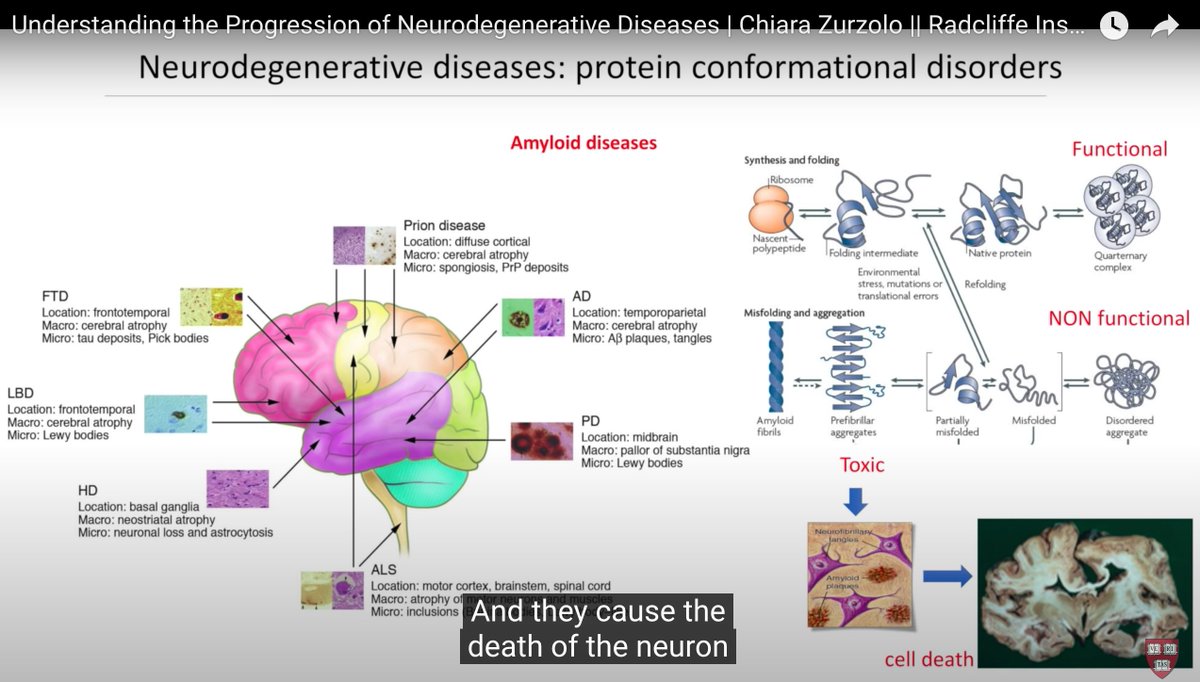
32/ “Neurodegenerative diseases are largely characterized by misfolding of proteins that accumulate in different areas of the brain." These aggregates are toxic, cause cell death, & are spread between cells by TNTs. -Dr. Chiara Zurzolo @institutpasteur
📽️
📽️
33/ MANY viruses including retroviruses, herpesviruses, orthomyxoviruses, & several others have been reported to trigger the formation of TNTs or TNT-like structures in infected cells and use these structures to efficiently spread to uninfected cells.
journals.asm.org/doi/10.1128/JV…
journals.asm.org/doi/10.1128/JV…
34/ In July 2022, Dr. Chiara Zurzolo and her team @institutpasteur actually showed TNT formation in culture and the ability to spread #SARSCoV2. However, this work did not use primary neurons.
PAPER: science.org/doi/10.1126/sc…
PAPER: science.org/doi/10.1126/sc…


35/ In 2021, she and her group @institutpasteur discovered that patient-derived glioblastoma (GBM) stem cells grown in 2D culture & 3D-tumor organoids, formed functional TNTs which allowed transfer of mitochondria. GBM is agressive and can relapse after tx
pubmed.ncbi.nlm.nih.gov/33245115/
pubmed.ncbi.nlm.nih.gov/33245115/
36/ This mechanism is seen in malignancy & infections. It is QUITE conserved from an evolutionary perspective.
THIS is why impact on neuronal function & looking at brain activity via objective tools like @wavimedical and @righteyeinsight are critical.
THIS is why impact on neuronal function & looking at brain activity via objective tools like @wavimedical and @righteyeinsight are critical.
https://twitter.com/ErinSandersNP/status/1629568121894846464?s=20
37/ This protein accumulation and failure to clear infection or malignancy out of the host cells, may also be why we see cognitive function changes and why objective tools like @BrainCheck & @CNSVitalSigns are imperative to quantify. cc @WesElyMD @RuhoyMD
https://twitter.com/ErinSandersNP/status/1630640042950443025?s=20
38/ We need to pair this objective data w/ subjective patient symptoms particularly in relation to debilitating post exertional malaise (PEM) from cognitive exertion, as well as physical & emotional exertion. [see DSQ-PEM or PAQ @sunsopeningband]
researchgate.net/publication/35…
researchgate.net/publication/35…
39/ I'm becoming more and more aware of how very wise our bodies are, if we will just actually listen to them
That maybe our bodies aren't broken, but functioning exactly as they were designed & telling us that something is very wrong and we're missing it
That maybe our bodies aren't broken, but functioning exactly as they were designed & telling us that something is very wrong and we're missing it
https://twitter.com/ErinSandersNP/status/1658325785420214272?s=20
40/ Dr. Chiara Zurzolo Presentation:
And hear me out, because of what we know about MS and EBV, or AD and HSV, what if...what is driving the protein propagation, at least in subsets of people, *is* actually these common infections that spread via TNT?
And hear me out, because of what we know about MS and EBV, or AD and HSV, what if...what is driving the protein propagation, at least in subsets of people, *is* actually these common infections that spread via TNT?
41/ Seriously, you should watch the whole thing.
• • •
Missing some Tweet in this thread? You can try to
force a refresh