
We found unique breast immune cells in the duct walls – ductal macrophages. They remove dying cells, help remodelling + are key players in cancer.
How did we get from this first sighting to finding their identity and function? Follow this thread!
bit.ly/35o2XSj
1/n
How did we get from this first sighting to finding their identity and function? Follow this thread!
bit.ly/35o2XSj
1/n
The breast contains mammary ducts surrounded by fat, blood vessels, immune + other cells.
We use 3D imaging to investigate the complex relationships b/w these cells to better understand disease.
#microscopy #mammarygland
2/n
We use 3D imaging to investigate the complex relationships b/w these cells to better understand disease.
#microscopy #mammarygland
2/n
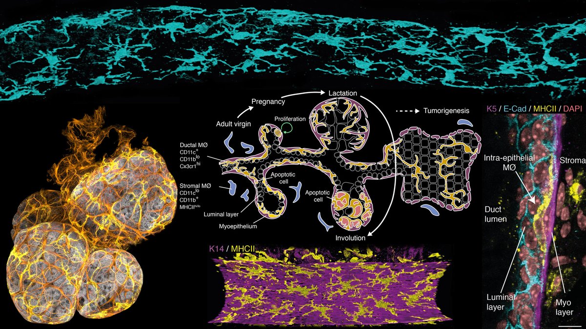
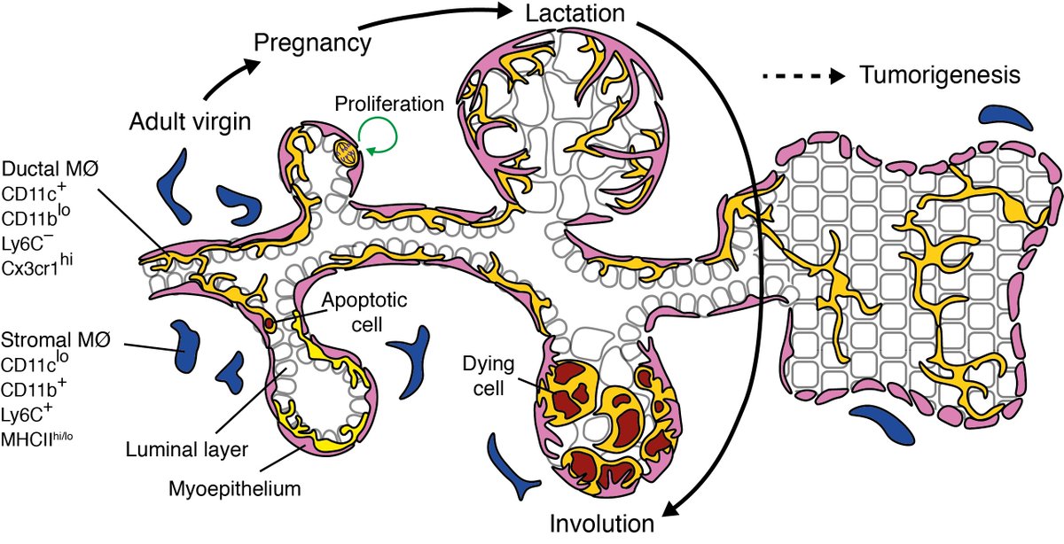
The ducts form a tree that branches through the breast and blooms and recedes in pregnancy and weaning. These are precancerous mouse ducts, alveoli in lactation and a summary of development in mice.
3/n
3/n


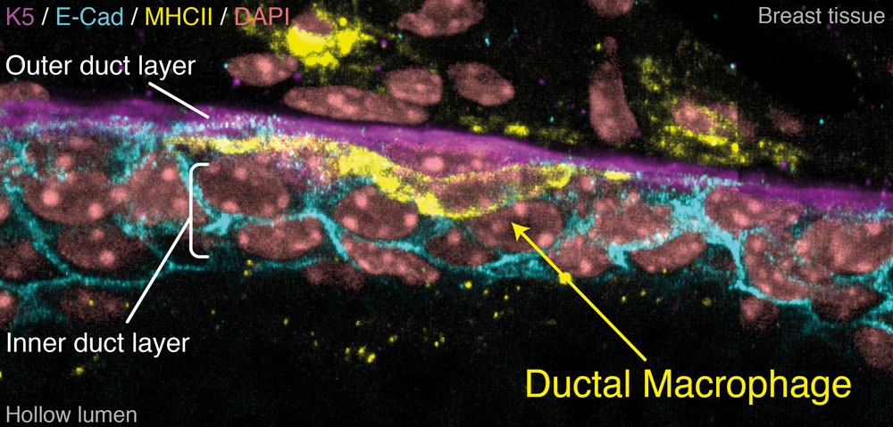
Mammary ducts have a two-layered epithelial wall: An outer layer of elongated, muscle-like cells (myoepithelial, Keratin 5) and an inner layer of 'luminal' cells (E-Cadherin), some of which make milk in lactation.
4/n
4/n
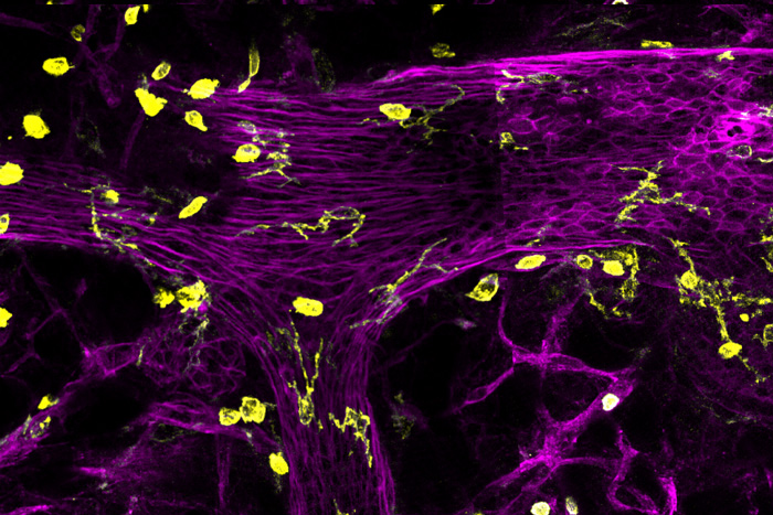
Going back 5 years… I started my PhD with Jane Visvader, Anne Rios and Geoff Lindeman, imaging different cell types to see what might interact with the ducts. Here are some nerve and immune cells. Some immune cells are lying on the ducts but they're hard to pick out.
5/n
5/n
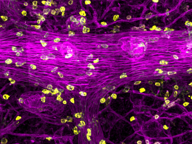
Looking closer in 3D (left) and in image slices (right)… there are immune cells INSIDE the duct! Very excited at this point but we had no idea what they were. We got lots of advice from immunologists, especially @SMuellerLab.
6/n
6/n

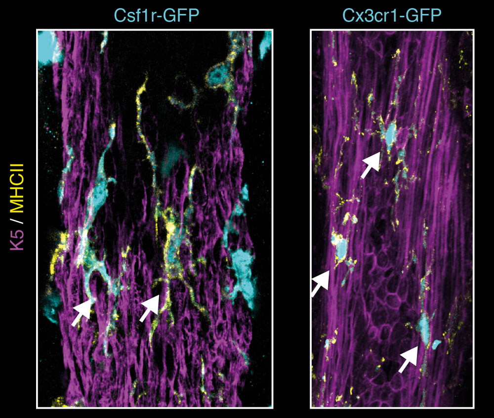
We imaged fluorescent gene reporters and found that they were Csf1r+ and Cx3cr1+, typical of immune cells called macrophages (MØs). These specialize in eating dead and infected cells and pathogens. #immunology
7/n
7/n
They are sandwiched between the layers of the duct wall!
They're so common inside the ducts that they form a network over the ducts.
We called them ductal macrophages, or 'DMs'.
8/n
They're so common inside the ducts that they form a network over the ducts.
We called them ductal macrophages, or 'DMs'.
8/n


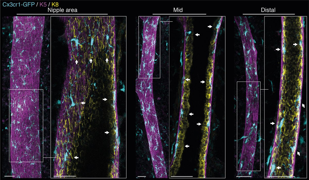
They cover the entire tree – near the nipple, in the major ducts and at the duct ends, always with the same regular spacing. This is all in mice, but it looks like they're in human ducts as well.
9/n
9/n


I developed a technique to image mammary ducts in vivo (with help from @SMuellerLab) and used this to watch the interaction between DMs (yellow) and the duct (purple). This was sooo cool to see. They touch all duct cells every few hours.
#intravital #microscopy
10/n
#intravital #microscopy
10/n
They phagocytose (eat) dying cells and we also filmed their response to targeted laser damage to the duct.
11/n
11/n
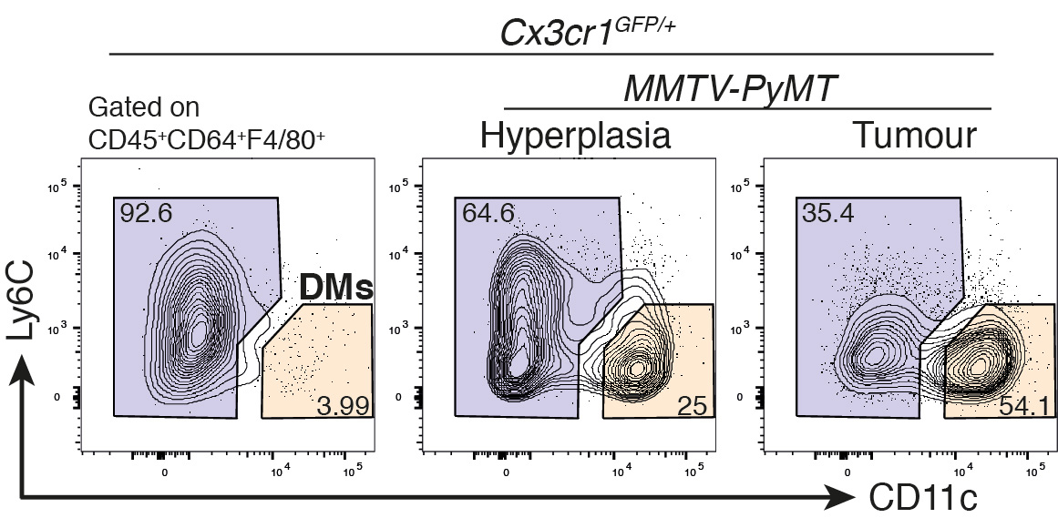
If only we could sort and isolate them to find out more... We needed unique markers for this, so I looked by imaging. I eventually found that they are CD11c+ and CD11b–, a bit unusual for MØs.
12/n
12/n

This translated perfectly into flow cytometry – we saw a small but very nice population of DMs!
13/n
13/n

To double check these were really the DMs, we looked at cells from just the fat without ducts… no DMs at all! And there were more when we enriched for ducts.
So, DMs only exist within the mammary ducts.
14/n
So, DMs only exist within the mammary ducts.
14/n

Now we could isolate them for RNA-seq. This showed they are really different to other breast immune cells. It also revealed Notch activation in DMs and too many unique genes to know what to do with.
15/n
15/n


Now that we had nailed these guys down, we could really get into the biology. We made bone-marrow chimeras, which show us how long it takes for cells to be replaced: DMs live in the ducts for about 5 months!
(MØ ppl see 👇 for origin 😉)
16/n
(MØ ppl see 👇 for origin 😉)
16/n

We could also track them throughout breast development. This is log scale, % of total cells… look at them go through pregnancy and into lactation!! (Inv = involution)
17/n
17/n

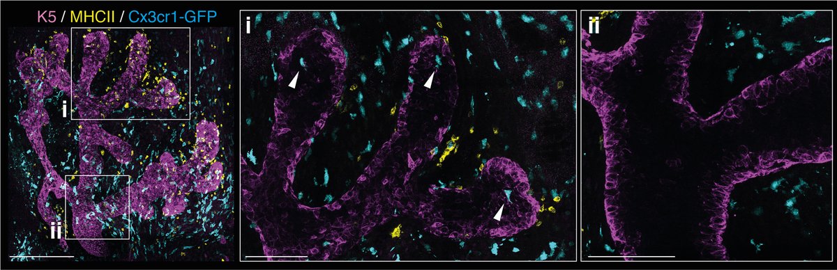
Where are they in lactation? DMs (yellow, MHCII) snuggle up with the myoepithelial cells (purple, K5) that wrap around alveoli. They're still between the two layers and cover the gland with similar density. Pink = F-actin.
#microscopy
18/n
#microscopy
18/n
After weaning, all milk-producing (alveolar) cells die and the alveoli collapse. This is called involution. Imaging this stage showed that DMs fill the alveoli. Are they eating the dying cells?
19/n
19/n

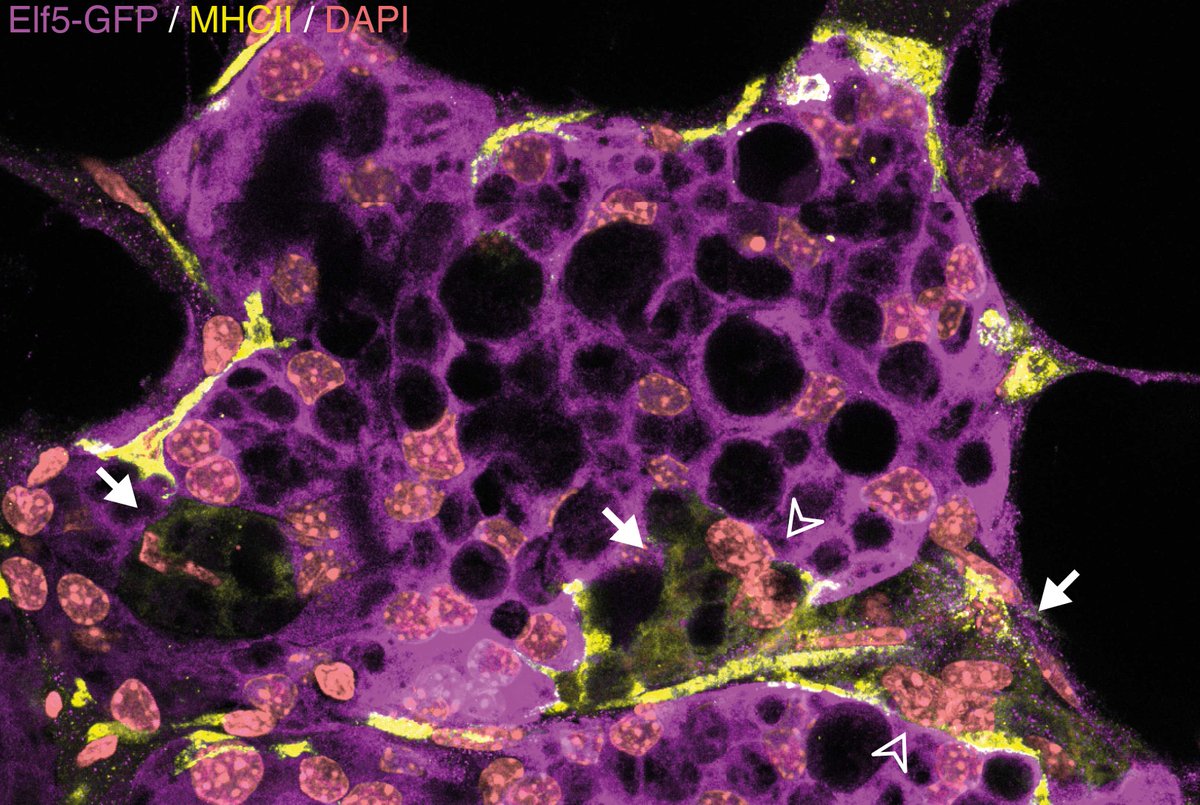
When I imaged fluorescent alveolar cells (Elf5-GFP) before the alveoli had collapsed, I could see some cells with DMs wrapping around them.
20/n
20/n

Filming in vivo showed DMs taking up cells with the fluorescent signal.
So DMs eat the dying cells, but are they necessary for remodelling? This process is like wound healing, is associated with cancer and prepares the gland for new pregnancies.
21/n
So DMs eat the dying cells, but are they necessary for remodelling? This process is like wound healing, is associated with cancer and prepares the gland for new pregnancies.
21/n
Removing DMs during involution (by CD11c-DTR or anti-Csf1r) slowed alveolar collapse (bigger lumen) and left dying cells (blue) uneaten. Now we know DMs help the breast return to normal and that other cells can't do the job.
22/n
22/n

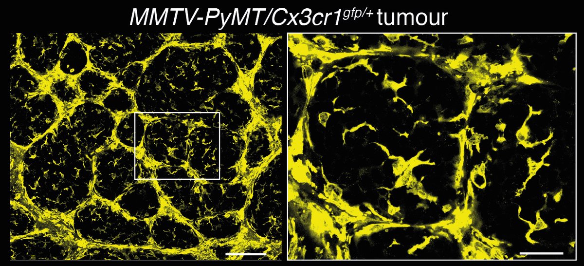
Focusing on disease: #breastcancer MØs (#TAMs) limit the effectiveness of cancer treatment. They are different to normal breast MØs but it's not clear why…
…but they are like DMs!
23/n
…but they are like DMs!
23/n
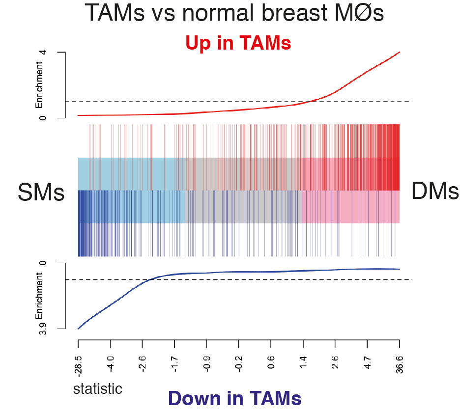
So MØs are probably educated by similar factors in breast cancer and in the breast ducts. Will this help us to target them to improve treatment? What role do they play in early stage cancer? Interesting questions!
24/n
24/n
In summary, we discovered a new population of tissue-resident macrophages in breast ducts!
Please welcome DMs into the #macrophage family 🤗
Thanks to the whole, incredible team!
bit.ly/mammaryDMs bit.ly/35o2XSj
25/n
Please welcome DMs into the #macrophage family 🤗
Thanks to the whole, incredible team!
bit.ly/mammaryDMs bit.ly/35o2XSj
25/n
Here's a quick summary of what we found, but there are more interesting points to discuss below. Especially for #macrophage enthusiasts.
26/n
26/n
Where do DMs come from?? We found out while on an imaging meeting trip to Singapore (thanks to @lai9uan) only 2 weeks after the Ms4a3 pre-print from @FGinhoux and co.
Spontaneous collaboration through #ScienceTwitter!
27/n
Spontaneous collaboration through #ScienceTwitter!
27/n
We smashed out the experiments in a week:
Ms4a3-Cre/tdTom only labels bone-marrow-derived MØs, not embryonic. In the tiny prepubertal gland, DMs are embryonic, but as they increase along with duct growth they are bone-marrow-derived.
28/n
Ms4a3-Cre/tdTom only labels bone-marrow-derived MØs, not embryonic. In the tiny prepubertal gland, DMs are embryonic, but as they increase along with duct growth they are bone-marrow-derived.
28/n

Where are these embryonic DM precursors?
I imaged whole embryonic glands and found one #macrophage inside each duct tip – maybe these can generate all DMs until puberty? They only turn on MHCII at 1 week. #microscopy
29/n
I imaged whole embryonic glands and found one #macrophage inside each duct tip – maybe these can generate all DMs until puberty? They only turn on MHCII at 1 week. #microscopy
29/n
Do DMs proliferate? Yes!
Not in the steady state, but to drive expansion in pregnancy (~5% EdU+).
30/n
Not in the steady state, but to drive expansion in pregnancy (~5% EdU+).
30/n

The breast completely transforms throughout life. DMs are always there – do they change as well?
Yes, many genes change in expression during pregnancy and lactation, but after involution, they return to the same resting state.
31/n
Yes, many genes change in expression during pregnancy and lactation, but after involution, they return to the same resting state.
31/n

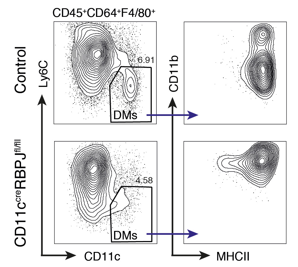
Finally, we followed up on the Notch signature by disrupting Notch signalling in DMs. After deleting the Notch effector RBPJ, DMs were still present in the ducts but failed to down-regulate CD11b. Looks like Notch directs DM maturation.
32/n
32/n

Very interesting in light of Notch's role in MØ/mammary stem cells signalling doi.org/10.1126/scienc…
and breast cancer MØs doi.org/10.1126/scienc…, from @RAFranklin_
33/n
and breast cancer MØs doi.org/10.1126/scienc…, from @RAFranklin_
33/n
If you made it the whole way… WOW! That was long 😅
You get a medal 🏅
Check out my blog on the paper bit.ly/mammaryDMs
or the full paper bit.ly/35o2XSj. Free access here rdcu.be/b3PJY.
34/n
You get a medal 🏅
Check out my blog on the paper bit.ly/mammaryDMs
or the full paper bit.ly/35o2XSj. Free access here rdcu.be/b3PJY.
34/n
Thanks for joining the thread!
Please ask questions. I have so much time and I'm stuck at home 😁
35/the end *DMs waving
Please ask questions. I have so much time and I'm stuck at home 😁
35/the end *DMs waving
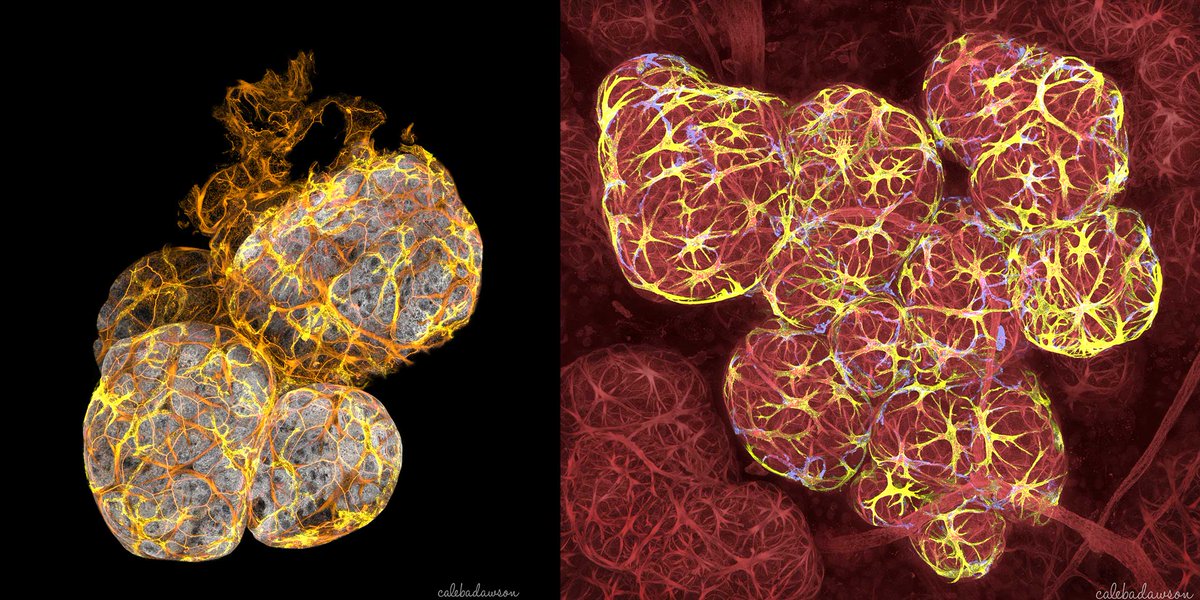
So happy to have my image on the cover!
What a great way to cap off this journey.
Can't wait to have a hard copy (I might frame it haha ☺️)
What a great way to cap off this journey.
Can't wait to have a hard copy (I might frame it haha ☺️)
@threadreaderapp unroll
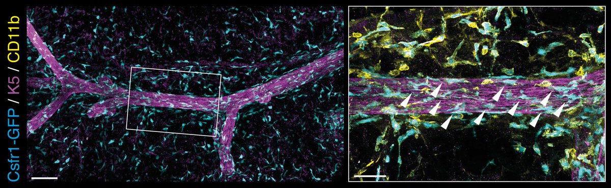
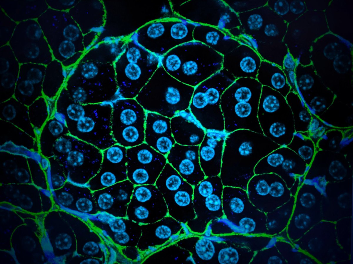
I missed out this nice 3D image with macrophages in blue, showing that the breast ductal macrophages are CD11b–
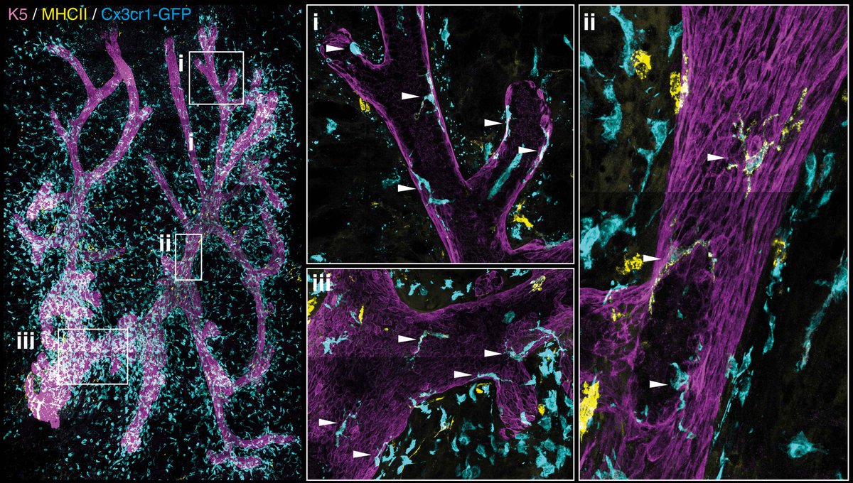
Imaging of a whole mammary gland at 4 days of age – there are more macrophages at the duct tips (i) and a few creeping down towards the nipple (iii). High-resolution to zoom in.

In a 1-week mammary gland, they're more spread over the tree and starting to look more like mature DMs.
#microscopy #macrophages #3Dimaging #mammarygland
#microscopy #macrophages #3Dimaging #mammarygland
More breast macrophages (yellow) eating milk-producing cells (purple, left), because it's amazing. Milk-producing cells are huge and binucleated (right). If you zoom in to the first image you can see the 2 nuclei inside the macrophages (hollow arrows) 😎 #cellbiology
Free access to the paper here: rdcu.be/b3PJY
Tissue-resident ductal macrophages survey the mammary epithelium and facilitate tissue remodelling @NatureCellBio #SciArt #microscopy #openaccess
Tissue-resident ductal macrophages survey the mammary epithelium and facilitate tissue remodelling @NatureCellBio #SciArt #microscopy #openaccess


