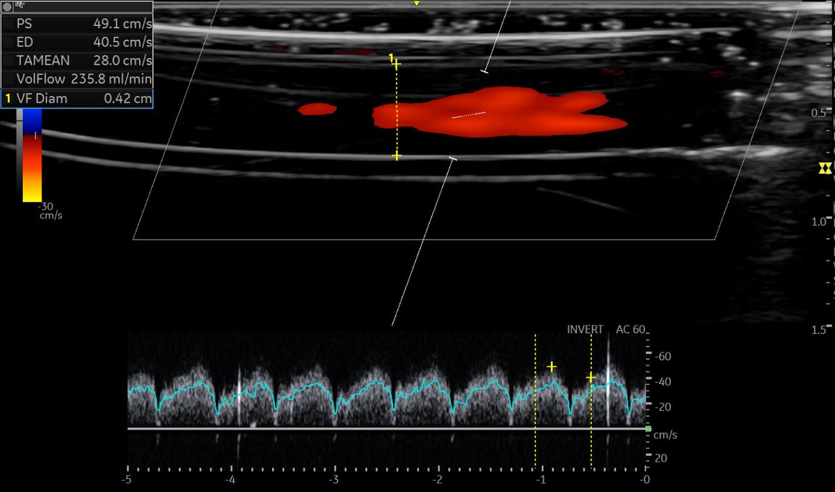
Stimulated by the enthusiasm of #MedEd student and #nephrology fellow, did a small experiment to see how well #POCUS -determined blood flow in the continuous renal replacement therapy (CRRT) circuit correlates with the actual no. 1/ First, got a color #Doppler img. of the tube 👇
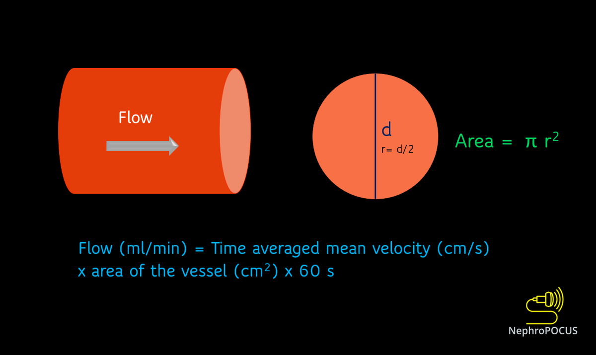
2/ How do you calculate flow? It is the same principle that we use to determine flow rate in an arteriovenous fistula 👇 #POCUS
3/ 2nd step: measured the diameter of the tube (double-lined structure) - 0.42 cm
4/ Now we get a pulsed wave Doppler tracing of the blood flow and the US machine does rest of the job - calculates the product of time-averaged mean velocity (TA Mean) & the area.
Result = 235.8 ml/min...pretty accurate considering the slippery tube & my hand full of gel! #POCUS
• • •
Missing some Tweet in this thread? You can try to
force a refresh
#Nephrology#POCUS case of the day:
What do you think is this anechoic structure adjacent to liver?
See thread 👇 for the answer and more images. #MedEd#IMPOCUS
Let's start with a poll before seeing other images: ☝️? #POCUS
The answer is right renal cyst. Note how the kidney appears with fanning the probe. #POCUS
Is rest of the kidney normal? doesn't appear to be...🤔
Looks D-only but could this be S-wave? (or a delayed D? Pt has Afib and predisposed to having smaller S but wondering if there is some S-D fusion here)
Renal #POCUS in a #dialysis patient: Looks fine at first glance (thin parenchyma as expected) but fanning anteriorly reveals a large cyst, which would have been missed otherwise. Labeled images and notes in thread 👇 #Nephrology#MedEd#IMPOCUS
Labeled first image #POCUS
Note: peri-renal fat (usually prominent in ESRD patients) can be confused with free fluid.
Labeled second image #POCUS (obtained with phased array probe)
Note: EKG is not needed 😀
#Nephrology consult for worsening creatinine in the context of diuresis in a patient with #heartfailure
Documented weight 3 kg ⬇️ than the previous day. No accurate urine output. Pt feels OK (has some unrelated issue).
Question: over diuresis? #POCUS#MedEd#VExUS see thread 👇
So, there is decreased LV systolic function and obviously diastolic dysfunction. Bi atrial enlargement. No significant pericardial effusion (there was trace on PLAX).
Next step in the hemodynamic circuit #POCUS assessment?
btw repeat weight was similar.
Next: IVC #POCUS assessment to assess right atrial pressure.
Here it is 👇
Big and plethoric (take it as 15 mmHg in a spontaneously breathing person - we get RVSP of ~41 mmHg based on the above tricuspid gradient)
#POCUS Intra-renal vein Doppler waveform looking exactly like that of hepatic vein! #VExUS obtained from a #nephrology clinic patient with pulmonary hypertension. #MedEd