2/7...CN 3,6,V1 travel between two dural layers in the lateral wall of the CS. V2 often travels in the the inferolateral wall of the CS (sometimes inferior to the CS). Cranial nerve 6 floats freely in the CS (why CS pathology often selectively affects CN6).
3/7...Note that: postganglionic sympathetic inputs to the orbit (originating from the sup cervical ganglion) ascend with the ICA, branch off the ICA in the CS, and then join branches of CN3 (to sup tarsal muscle) and V1 (pupillary dilation).
4/7...CNs 3 and V1 can usually be easily seen with imaging in the lateral wall of the CS, while the tiny CN4 is rarely identified. CN6 can sometimes be faintly seen adj. to the ICA in the CS. Each will extend anteriorly to exit the cranial cavity via the sup orbital fissure.
5/7...CN V2 at the inferior edge of the sinus will travel anteriorly to exit via the foramen rotundum towards the pterygopalatine fossa.
6/7...CN V3 travels briefly along the floor of the middle cranial fossa inferior to the cavernous sinus, exiting the intracranial cavity inferiorly through the foramen ovale.
7/7...Last point: although some controversy and ambiguity persist, the medial wall of the CS is likely not a true dural layer (despite most diagrams, including mine), but rather a weak fibrous border. This predisposes the CS to invasion by pituitary adenoma.
From neurosurgeons at Pitt--fine details of the medial wall of CS--which they find is in fact a single dural layer splitting from double layer sellar floor--plus technique for dissection of parasellar ligaments. thanks for the reference @DrCohenCohen! thejns.org/view/journals/…
• • •
Missing some Tweet in this thread? You can try to
force a refresh
2/5...The STN is functionally a node within the basal ganglia (BG) INDIRECT LOOP. STN contains excitatory glutaminergic neuronsâžžoutput to the GABA neurons of GPi, which in turn have inhibitory effect on thalamic outputs to the motor cortex.
3/5...Loss of nigrostriatal input in Parkinsons Dzâžžincreased inhibitory output from GPiâžždecreased thalamic stimulation of the motor cortex (through both direct and indirect loop circuits). Makes sense that DBS treatments were initially directed at disrupting activity in GPi.
2/6...Meckel’s cave (MC) is open to the subarachnoid space at its posterior margin (and is therefore filled with CSF). The trigeminal (Gasserion) ganglion lives in MC. Superiomedial to MC (and sharing a dural border), is the cavernous sinus. #radres#neurorad
3/6...Coursing anteriorly, the V1 and V2 branches of CNV exit MC to travel within the lateral wall of the cavernous sinus. V3 courses inferiorly to exit the middle cranial fossa through foramen ovale, without involving the cavernous sinus.
2/8..The HC: complex structure of limbic system; encodes memories from short->long term (also involved in pattern recognition,memory encoding & association, working memory,spatial nav, emotional behavior,awareness of conscious knowledge). Am I missing anything neuroscientists?
3/8...Anatomically: the HC is made of the cornu ammonis (CA) and dentate gyrus (DG); hippocampal formation also includes the subiculum and entorhinal cortex (EC). Can also be divided into head/body/tail. Supplied primarily by the PCA, and variably by anterior choroidal artery.
ICA segments: C1 (cervical) becomes C2 (petrous) in the carotid canal of the petrous bone. Becomes C3 (lacerum) as it exits the carotid canal above the foramen lacerum. Becomes C4 (cavernous segment above the petrolingual ligament through the cavernous sinus.
...C5 (clinoid) above proximal to the distal dural ring. C6 (ophthalmic) is truly intracranial. C7 (communicating) distal to the Pcomm. Alternative segmentation schemes include C1-C4 (cervical, petrous, cavernous, supraclinoid/terminal)
The AC runs across the midline in front of the anterior columns of the fornix, behind the basal forebrain and beneath the anterior limb internal capsule and basal ganglia, surrounded by the bed nucleus of the stria terminalis.
The AC connects areas of the bilateral temporal poles and orbitofrontal cortex. Function is not entirely understood but it is thought to be important in the olfactory pathway and pain sensation, among other things.
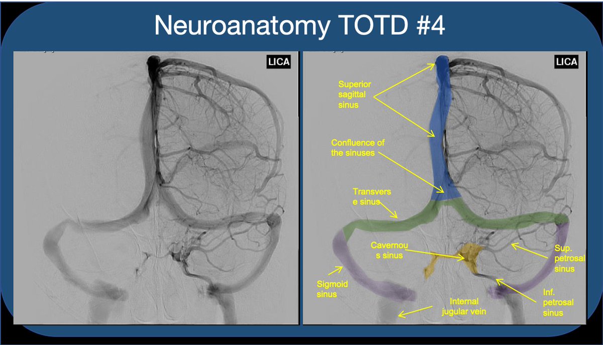
Neuroanatomy TOTD #4 1/5 Answer: The orange structure is the cavernous sinus (CS), a paired dura-lined venous cavity on either side of the sella. The sinuses are split into numerous “caves” by fibrous septae (hence the name). #neuroanatomy#neurorad#medtwitter#neuroanatomyTOTD
2/5 Note that the paired sinuses are often variably connected by “intercavernous sinuses”. While the CS is often taught for its relationship to the ICA and cranial nerves, I find that medical students and residents rarely understand the flow of blood through the sinus.
3/5 The CS receives blood from sup. and inf. ophthalmic veins draining the orbit--This is how a facial/orbital infection spreads intracranially to CS (classic #usmle ?). The sphenoparietal sinus as well as the superficial middle and inferieor cerebral veins also feed into the CS.