Neuroanatomy TOTD #11🧵
➡️intracanalicular facial nerve. IAC cross section➡️4 nerves; ant-sup➡️facial, ant-inf➡️cochlear (7-up/coke-down). Post nerves➡️sup&inf vestibular #medtwitter #meded #FOAMed #FOAMrad #neurorad #radiology #radres #neurology #neurosurgery #neuroanatomy
1/11
Facial motor nucleus is in the pontine tegmentum; axons loop dorsally around the abducens nucleus, then course anterolaterally, exiting the brainstem at the CPA.
2/11
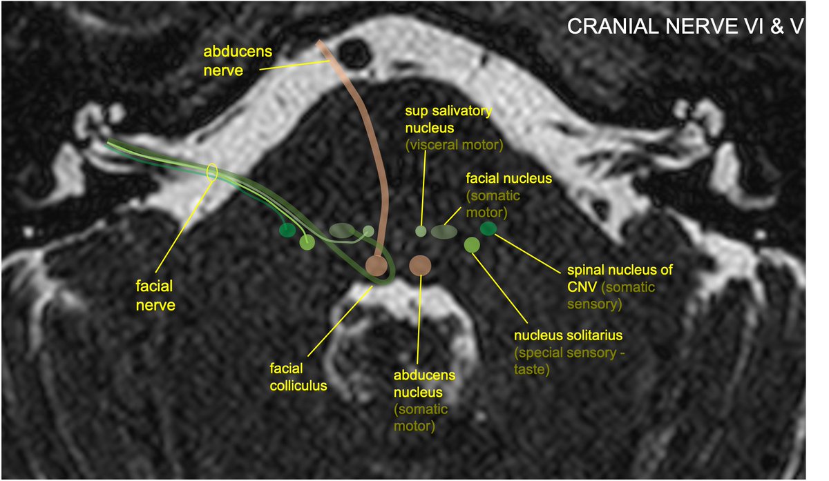
The nervus intermedius (NI) is composed of preganglionic parasympathetic fibers w/cell bodies in the sup salivatory nucleus, taste fibers w/cell bodies in the nucleus solitarius, and somatic sensory input from EAC/external ear, w/cell bodies in the spinal nucleus of CNV.

3/11
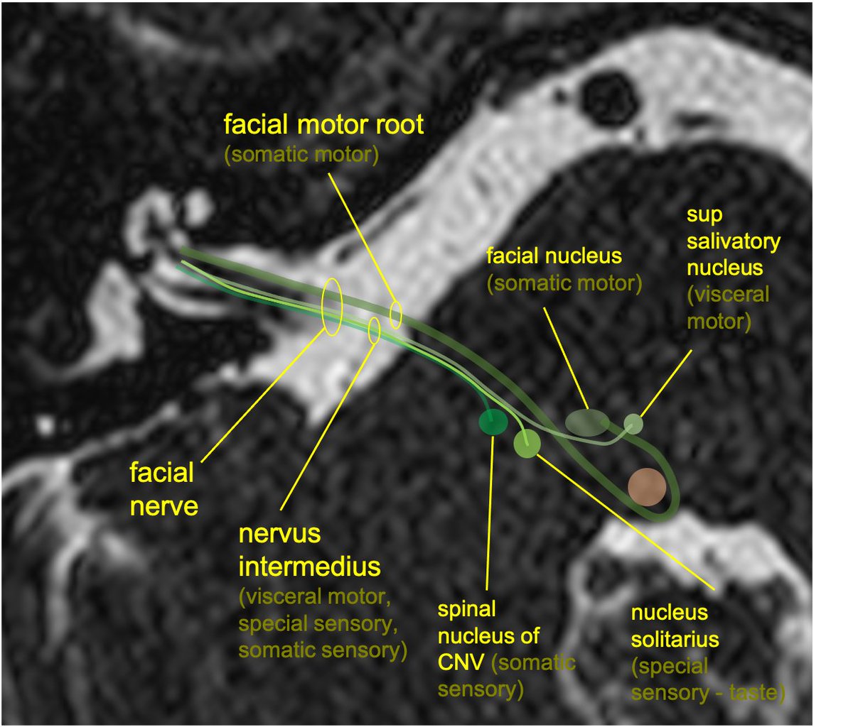
Although NI and the motor components of CN7 are anatomically separate, they are generally not distinguished on imaging of the IAC.

4/11
Facial nerve is separated into 4 intratemporal segments (intracanalicular, labyrinthine, tympanic, mastoid). Cisternal is from the CPA to the internal auditory meatus. Intracanalicular within the IAC. Labarynthine is from the fundus of the IAC to the geniculate ganglion.

5/11
The greater superficial petrosal nerve (GSPN) arises from geniculate ganglion w/preganglionic parasymp fibers (and some taste/sensation) along floor of middle cranial fossa, through pterygoid canal, to pterygopalatine fossa➡️lacrimal gland(TOTD #1) ()

6/11
The tympanic segment starts at the sharp turn of the geniculate ganglion and runs horizontally/posterolaterally along the medial margin of the middle ear. (There are no branches of the tympanic segment).

7/11
At the post middle ear, the mastoid segment begins➡️turns inferiorly through the facial canal in the mastoid, giving off 3 branches: nerve of stapedius, chorda tympani, and sensory auricular branch.

8/11
The chorda tympani includes taste fibers to the ant 2/3 of the tongue, as well as preganglionic parasympathetic fibers which extend to the submandibular ganglion. The ch tymp traverses the middle ear and eventually joins with the lingual nerve (CNV) on its way to the tongue

9/11
CN7 exits bony facial canal through stylomastoid foramen➡️gives off 3 motor branches: post auricular, digastric, stylohyoid. Traverses parotid gland, divides into 5 branches: temporal, zygomatic, buccal, marginal mandibular, cervical (mnemonic: To Zanzibar By Motor Car).

10/11
Relevant path: Bell’s palsy (may be HSV reactiv in geniculate ganglion); variable effects on CN7: facial palsy, hyperacusis (stapedius), pain at jaw/ear (sensory auricular), loss of taste (ch tymp), changes in tear (GSPN) and saliva production (ch tymp)
#radiopaedia case

11/11

• • •

Missing some Tweet in this thread? You can try to force a refresh
 

Keep Current with Aaron Rutman, MD

Aaron Rutman, MD Profile picture

Stay in touch and get notified when new unrolls are available from this author!

Read all threads

This Thread may be Removed Anytime!

PDF

Twitter may remove this content at anytime! Save it as PDF for later use!

Try unrolling a thread yourself!

how to unroll video
  1. Follow @ThreadReaderApp to mention us!

  2. From a Twitter thread mention us with a keyword "unroll"
@threadreaderapp unroll

Practice here first or read more on our help page!

More from @aaronrutman

30 Oct
Neuroanatomy TOTD #10🧵 
1/5 Small gray matter structure at the junction of the thalamus and midbrain is the subthalamic nucleus (STN).

#meded #FOAMed #FOAMrad #medtwitter #medstudents #radiology #neurorad #radres #neurology #neurosurgery #neuroanatomy #neuroanatomyTOTD
2/5...The STN is functionally a node within the basal ganglia (BG) INDIRECT LOOP. STN contains excitatory glutaminergic neurons➞output to the GABA neurons of GPi, which in turn have inhibitory effect on thalamic outputs to the motor cortex.
3/5...Loss of nigrostriatal input in Parkinsons Dz➞increased inhibitory output from GPi➞decreased thalamic stimulation of the motor cortex (through both direct and indirect loop circuits). Makes sense that DBS treatments were initially directed at disrupting activity in GPi.
Read 5 tweets
23 Oct
Neuroanatomy TOTD #9🧵
1/6 The trigeminal n. courses anteriorly➡️prepontine cistern➡️into Meckel’s cave (green), which lies at the medial floor of middle cranial fossa at the petrous apex.
#meded #FOAMed #FOAMrad #neurorad #neurology #neurosurgery #neuroanatomy #neuroanatomyTOTD Image
2/6...Meckel’s cave (MC) is open to the subarachnoid space at its posterior margin (and is therefore filled with CSF). The trigeminal (Gasserion) ganglion lives in MC. Superiomedial to MC (and sharing a dural border), is the cavernous sinus. #radres #neurorad Image
3/6...Coursing anteriorly, the V1 and V2 branches of CNV exit MC to travel within the lateral wall of the cavernous sinus. V3 courses inferiorly to exit the middle cranial fossa through foramen ovale, without involving the cavernous sinus. Image
Read 6 tweets
13 Oct
Neuroanatomy TOTD #8🧵
1/8 MRI shows the hippocampal formation; the hippocampus(HC) is a ridge of archicortex gray matter at floor of lat ventricle, in the med temp lobe #FOAMed #FOAMrad #radres #neurorad #neuroscience #medtwitter #radiology #neurology #neurosurgery #neuroanatomy Image
2/8..The HC: complex structure of limbic system; encodes memories from short->long term (also involved in pattern recognition,memory encoding & association, working memory,spatial nav, emotional behavior,awareness of conscious knowledge). Am I missing anything neuroscientists?
3/8...Anatomically: the HC is made of the cornu ammonis (CA) and dentate gyrus (DG); hippocampal formation also includes the subiculum and entorhinal cortex (EC). Can also be divided into head/body/tail. Supplied primarily by the PCA, and variably by anterior choroidal artery. Image
Read 9 tweets
6 Oct
Neuroanatomy TOTD #7🧵

1/7 The cavernous sinuses (CS) are outlined on either side of the sella. The ICA & cranial nerves 3,4,6,V1,V2 travel through the CS.

#meded #FOAMed #FOAMrad #radres #neurorad #medtwitter #radiology #neurology #neurosurgery #neuroanatomy #neuroanatomyTOTD Image
2/7...CN 3,6,V1 travel between two dural layers in the lateral wall of the CS. V2 often travels in the the inferolateral wall of the CS (sometimes inferior to the CS). Cranial nerve 6 floats freely in the CS (why CS pathology often selectively affects CN6). Image
3/7...Note that: postganglionic sympathetic inputs to the orbit (originating from the sup cervical ganglion) ascend with the ICA, branch off the ICA in the CS, and then join branches of CN3 (to sup tarsal muscle) and V1 (pupillary dilation).
Read 8 tweets
29 Sep
Neuroanatomy TOTD #6
a) Ant choroidal artery infarct, involving the post limb of the internal capsule
b) Sup hypophyseal artery aneurysm—medially directed from the supraclinoid ICA
#meded #FOAMed #FOAMrad #radres #neurorad #radiology #neurosurgery #neuroanatomy #neuroanatomyTOTD ImageImage
ICA segments: C1 (cervical) becomes C2 (petrous) in the carotid canal of the petrous bone. Becomes C3 (lacerum) as it exits the carotid canal above the foramen lacerum. Becomes C4 (cavernous segment above the petrolingual ligament through the cavernous sinus. Image
...C5 (clinoid) above proximal to the distal dural ring. C6 (ophthalmic) is truly intracranial. C7 (communicating) distal to the Pcomm. Alternative segmentation schemes include C1-C4 (cervical, petrous, cavernous, supraclinoid/terminal) Image
Read 5 tweets
24 Sep
Neuroanatomy TOTD #5

The indicated bundle is the anterior commissure (AC), located at the ant border of the 3rd ventricle, at the sup margin of the lamina terminalis.

#meded #FOAMed #FOAMrad #radres #neurorad #medtwitter #radiology #neurosurgery #neuroanatomy #neuroanatomyTOTD Image
The AC runs across the midline in front of the anterior columns of the fornix, behind the basal forebrain and beneath the anterior limb internal capsule and basal ganglia, surrounded by the bed nucleus of the stria terminalis.
The AC connects areas of the bilateral temporal poles and orbitofrontal cortex. Function is not entirely understood but it is thought to be important in the olfactory pathway and pain sensation, among other things.
Read 6 tweets

Did Thread Reader help you today?

Support us! We are indie developers!


This site is made by just two indie developers on a laptop doing marketing, support and development! Read more about the story.

Become a Premium Member ($3/month or $30/year) and get exclusive features!

Become Premium

Too expensive? Make a small donation by buying us coffee ($5) or help with server cost ($10)

Donate via Paypal Become our Patreon

Thank you for your support!

Follow Us on Twitter!