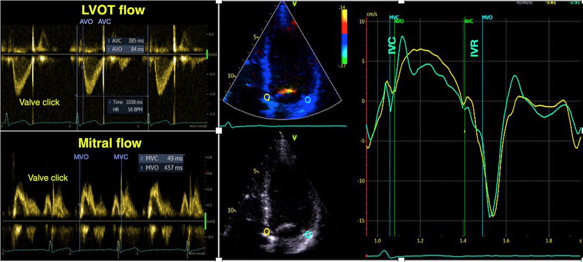
1/ During pre ejection, the vortex is seen to persist after MVC, and the septal part aligns with left ventricular outflow. This adds momentum and kinetic energy to the ejection flow.
2/ During ejection, however, the vortex seems to disappear, outflow more or less filling the whole apex, as flow in the lateral part is recruited by the rapid flow into the LVOT.
3/ At the same time, the AV plane with the closed mitral valve moves towards the apex. It seems that some of this recruited flow hits the mitral valve and is diverted back towards the apex, creating a basolateral momentum towards the apex again.
4/ -which after AVC leads into the IVC deformation creating a stronger momentum towards the apex before the start of early filling. Finding of a functional regional differential function (not "wasted work"), shows that Tau do not tell the whole truth about IVR.
1/ The intraventricular vortex fills the ventricle, and the downwards flow in the septal part, will close the anterior MV leaflet. This also isolates the vortex in the ventricle, which may conserve the kinetic energy in the vortex
2/ At the end of diastasis, the lateral part of the vortex, with apical flow, is aligned with the incoming inflow in atrial systole, adding momentum and kinetic energy to the inflow during atrial systole.
🧵In our paper Intraventricular Vector Flow Imaging with Blood Speckle Tracking in Adults: Feasibility, Normal Physiology and Mech… we use a new method, not only BST, and can be applied on adult probes. pubmed.ncbi.nlm.nih.gov/34620522/
The main aim was to investigate the normal adult, intraventricular blood flow throughout the whole cardiac cycle, to compare with pw and colour Doppler M-mode and wall mechanics. (2D images courtesy of AS Daae).
As tweeted before, during IVR, there is simultaneous shortening of the base and elongation of the apex, inducing a volume shift with intraventricular apical flow, imparting a momentum and kinetic energy towards apex before start of early filling. This is thus *not* "wasted work"
Apart from the physiological implications, what are the consequences of this study onlinelibrary.wiley.com/doi/epdf/10.11… for timing of valve openings and closures by tissue Doppler?
1/ Valve closures can be timed by tissue Doppler and mitral ring motion. However, only the septal motion will reliably show AVC.
2/ MVO is close to the END of the pre ejection spike. Timing MVC by the start of the pre ejection spike will result in an error of about 40 ms too early. Timing by the peak R wave will result in about the same error.
The negative velocity post ejection spike had an average duration of 35 ms, ending about 10 ms after AVC in the septum. Thus, this spike is not isovolumic relaxation, and the true IVR (AVC to MVO) is from the end of the spike to start of mitral flow.
2/ 22 healthy subjects, Valve openings and closures timed by Doppler flow, and transewfrred to Tissue Doppler recordings.
3/ Pre ejection velocities started 24.8 ms after start QRS, with a duration of 51.5 ms, ending about 11.5 ms after MVC. Thus, both electromechanical delay and pre ejection velocity occurs *before* onset of IVC, and are not a measure of IVC or Isovolumic acceleration.