Patient with flank pain, hematuria and significant leg edema

#POCUS 🧵

1/10
Lung Ultrasound #LUS 2/10
#IVC long axis 3/10
#IVC short axis + tilting 4/10
#echofirst PLAx + A4ch 5/10
Hepatic Vein Color Doppler 6/10
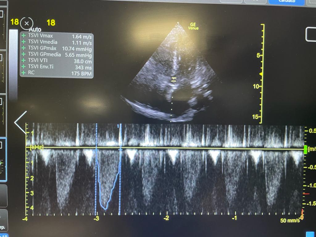
LVOT VTI = 13 7/10
Kidneys 8/10
Impression:
Dry lungs, IVC with intramural tumor extending to the right atrium. LV OK. No flow on IVC or HV. Low stroke volume. Normal right kidney (some free fluid). Left kidney tumor

9/10
There is severe leg edema with low preload (low stroke volume, no lung edema) and no flow in the IVC

This is Inferior Vena Cava Syndrome!

Cause is RCC invading the IVC and RA

Nice review here: doi.org/10.7326/0003-4…

END/

• • •

Missing some Tweet in this thread? You can try to force a refresh
 

Keep Current with Eduardo R Argaiz

Eduardo R Argaiz Profile picture

Stay in touch and get notified when new unrolls are available from this author!

Read all threads

This Thread may be Removed Anytime!

PDF

Twitter may remove this content at anytime! Save it as PDF for later use!

Try unrolling a thread yourself!

how to unroll video
  1. Follow @ThreadReaderApp to mention us!

  2. From a Twitter thread mention us with a keyword "unroll"
@threadreaderapp unroll

Practice here first or read more on our help page!

More from @ArgaizR

8 Dec
A tale of two hearts: Physiological observations on AV shunts and congestion 🧵

These are 2 patients on IHD I saw in the outpatient clinic

🔷 Both with severe venous congestion (#VExUS = 3)
🔷 Both with tortuous brachiocephalic AV fístula

1/11
What I found remarkable was the diametrically opposed effects of manual AVF compression on JVP! 🤯

🔴 Patient A: AVF Compression improves venous congestion
🔵 Patient B: AVF Compression worsens venous congestion

2/11
🔴 Patient A: SLE + Lupus Nefritis ➡️ ESRD in HD

#echofirst: Plethoric IVC, good LVEF, paradoxical septal motion, ventricular interdependence, severe RV/RA dilation, torrential TR

3/11
Read 13 tweets
28 Nov
Pt with advanced Cirrhosis

AKI, Oliguria + Encephalopathy

Initial treatment = Albumin + Lactulose enemas

🧂Na is now 158 (From Lactulose induced free water loss)

#POCUS ninja @tumleal noticed something was wrong! He got his probe and texted me

📱 #WhatsAppAKIConsult 1/5
#POCUS: Plethoric non collapsible IVC

Based on the clinical scenario, IVC, heart rate and Pulse Pressure...

What is the likely underlying pathophysiology? 2/5
@tumleal went ahead and confirmed his suspicion:

He 📲texted me this:

LVOT VTI = 38!

Some 🔢:

VTI of 38, assuming a LVOT diameter of 20 mm: Stroke Volume = 119 ml

119 ml x HR (93 bpm) = 11.1 L/min of CO!

This is High Output Heart Failure (Very common in Cirrhosis) 3/5
Read 5 tweets
21 Oct
Pt w right HF and high probability pulmonary hypertension

TAPSE 15 mm, TRVmax 4.1 m/s, paradoxical septal motion

Renal Venous Doppler 👇

According to doi.org/10.1161/JAHA.1…, Which curve color would best describe this patient's PH-related morbidity?

Poll and 🧵👇

1/6 ImageImage
Which curve in the Kaplan Meier Curve above best fits this particular patient?

2/6
The Renal Doppler shown 👆 looks like a biphasic pattern. This would mean the green curve 🟢

However there is a catch.....

3/6 Image
Read 6 tweets
28 Aug
One of my favorite and most intriguing causes of severe venous congestion (#VExUS = 3)

A 🧵on High Output Heart Failure (HOHF) 1/18
First, the index case:

Clip above shows hyperdynamic flow from the vena cava

#echofirst 👇: Very dilated and plethoric IVC, LV OK, Dilated RV, D sign

Overall: Increased Right heart filling pressures

2/18
IVC = 3.4 cm
Portal Vein = > 100% pulsatility
LVOT VTI = 26
TRVmax = 3.04 m/s

So we have:

Venous Congestion (IVC, Portal Vein)
High Cardiac Output (LVOT-VTI)
Pulmonary Hypertension (TRVmax)

3/18
Read 18 tweets
13 Jul
#AKIConsultSeries

👴 w Advanced Cirrhosis, hemorrhagic portal hypertension and hx of a heart block (w pacemaker).

Came to the ED w diarrhea 2/2 severe C. Diff.

Now in shock...

Initial resus with Norepinephrine 0.3 ug/kg/min, Crystalloid and albumin. Cr 3.8

1/7
18 hrs later, no renal improvement + oliguria.

MABP 70, CRT 2 seconds. 🧠 confused, + asterixis. No ascites or edema

Workup: hemodynamic AKI (⬆️SG, ⬇️UNa, ⬆️BUN/Cr, bland sediment)

Team wants to continue fluids, albumin and antibiotics... Dr. Harris, do you concur?

2/7
Obviously you are here for the #POCUS so here we go:

IVC: Plethoric (No subX window 2/2 intestinal air)
LV, RV: Relatively preserved systolic fx
Pacemaker lead seen causing important Tricuspid Regurgitation!

3/7
Read 7 tweets
3 May
AKI Consult: 👵 ➡️ ED with severe DKA. CT Abdomen and Chest to look for infectious trigger: negative. Tx with IV insulin and balanced crystalloid + 6 L with obvious improvement. Cr was 2.7

Remained oliguric, now in sudden shock with increasing NE dose (0.5 ucg/kg/min) 🚨 1/12
#POCUS Very hyper-dynamic🫀 with increased contractility and no RV dysfunction.

🔎 Look carefully at color of flow exiting the LV:

Aliasing (green color): This means ultrasound system is trying to image an event that is occurring faster than the sample rate

2/12
This means flow is fast. But how fast? Choose the CW doppler setting and find out!

In this case acceleration was almost 6 m/s!

Flow acceleration occurs in the setting of obstruction (similar to putting your finger on the hose exit)

So what is causing the obstruction? 3/12
Read 12 tweets

Did Thread Reader help you today?

Support us! We are indie developers!


This site is made by just two indie developers on a laptop doing marketing, support and development! Read more about the story.

Become a Premium Member ($3/month or $30/year) and get exclusive features!

Become Premium

Too expensive? Make a small donation by buying us coffee ($5) or help with server cost ($10)

Donate via Paypal

Or Donate anonymously using crypto!

Ethereum

0xfe58350B80634f60Fa6Dc149a72b4DFbc17D341E copy

Bitcoin

3ATGMxNzCUFzxpMCHL5sWSt4DVtS8UqXpi copy

Thank you for your support!

Follow Us on Twitter!

:(