1/
A #dermtwitter #tweetorial on...
#PEMPHIGUS VULGARIS!
Join me for a quick #thread on this autoimmune blistering disorder!
#MedEd #FOAMEd #medtwitter #MedStudentTwitter
A #dermtwitter #tweetorial on...
#PEMPHIGUS VULGARIS!
Join me for a quick #thread on this autoimmune blistering disorder!
#MedEd #FOAMEd #medtwitter #MedStudentTwitter

2/
Pemphigus vulgaris is where the patient's own antibodies target a Desmosomal protein, which leads to the keratinocytes coming apart.
I describe this to patients as a brick wall, where the mortar holding things together is getting dissolved.
Remember this?👇
Pemphigus vulgaris is where the patient's own antibodies target a Desmosomal protein, which leads to the keratinocytes coming apart.
I describe this to patients as a brick wall, where the mortar holding things together is getting dissolved.
Remember this?👇

3/
This is contrast to the Pemphigoid group of diseases, that target the hemidesmosome. In other words, remember that:
pemphiguS = Superficial (in the epidermis) (1)
pemphigoiD = Deep (below epidermis) (2)
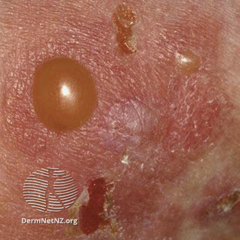
pemphigus = FLACCID blisters
pemphigoid = TENSE blisters
This is contrast to the Pemphigoid group of diseases, that target the hemidesmosome. In other words, remember that:
pemphiguS = Superficial (in the epidermis) (1)
pemphigoiD = Deep (below epidermis) (2)
pemphigus = FLACCID blisters
pemphigoid = TENSE blisters

4/
Remember that any tense blister can turn flaccid after some time, so always make sure you're evaluating a new lesion!
Don't forget in pemphigus you also see a + Nikolsky (negative in Pemphigoid)
What exactly is a Nikolsky?
Remember that any tense blister can turn flaccid after some time, so always make sure you're evaluating a new lesion!
Don't forget in pemphigus you also see a + Nikolsky (negative in Pemphigoid)
What exactly is a Nikolsky?
5/
A Nikolsky is positive if lateral pressure next to the blister recreates the blistering process!
If you push down on a blister and it spreads, that's an "indirect Nikolsky" or an Asboe-Hansen sign.
Here's a video curated by @grepmed of Asboe-Hansen!
img.grepmed.com/uploads/10423/…
A Nikolsky is positive if lateral pressure next to the blister recreates the blistering process!
If you push down on a blister and it spreads, that's an "indirect Nikolsky" or an Asboe-Hansen sign.
Here's a video curated by @grepmed of Asboe-Hansen!
img.grepmed.com/uploads/10423/…
6/
Okay, now that we've figured out what the exam looks like, let's talk distribution.
By definition, where should you expect to see erosions in pemphigus vulgaris?
Okay, now that we've figured out what the exam looks like, let's talk distribution.
By definition, where should you expect to see erosions in pemphigus vulgaris?
7/
The mouth should theoretically ALWAYS be involved!
That's because Pemphigus vulgaris is defined by antibodies targeting Desmoglein 3 (+/- Dsg 1).
Dsg 3 is preferentially expressed in the oropharynx, so the mouth should be involved!
pc: jaad.org/article/S0190-…
The mouth should theoretically ALWAYS be involved!
That's because Pemphigus vulgaris is defined by antibodies targeting Desmoglein 3 (+/- Dsg 1).
Dsg 3 is preferentially expressed in the oropharynx, so the mouth should be involved!
pc: jaad.org/article/S0190-…

8/
What about Dsg 1? That's expressed more on the skin, so if antibodies to Dsg1 are present, you're more likely to get skin disease.
This explain why pemphigus foliaceous & staph scalded skin (which both only effect Dsg 1), do NOT involve the mouth!
What about Dsg 1? That's expressed more on the skin, so if antibodies to Dsg1 are present, you're more likely to get skin disease.
This explain why pemphigus foliaceous & staph scalded skin (which both only effect Dsg 1), do NOT involve the mouth!
https://twitter.com/DrStevenTChen/status/1387730884544061442?s=20
9/
So how do we diagnose it?
Well, a biopsy for H+E is helpful, and when paired with a direct immunofluorescence, it can really confirm your diagnosis.
As you'd expect, a split in the epidermis is seen, with antibodies lighting up between the keratinocytes!
So how do we diagnose it?
Well, a biopsy for H+E is helpful, and when paired with a direct immunofluorescence, it can really confirm your diagnosis.
As you'd expect, a split in the epidermis is seen, with antibodies lighting up between the keratinocytes!


10/
Remember: direct immunofluorescence is the patient's skin!
You can also do an indirect immunofluorescence, where you take patient serum, and react it with a substrate rich in Dsg 3 (Monkey esophagus for those curious).
You can also check Dsg 3 and Dsg 1 antibody titers!
Remember: direct immunofluorescence is the patient's skin!
You can also do an indirect immunofluorescence, where you take patient serum, and react it with a substrate rich in Dsg 3 (Monkey esophagus for those curious).
You can also check Dsg 3 and Dsg 1 antibody titers!
11/
There are some reports of correlation of Dsg titers to severity of disease. For me, it doesn't really change how I manage the patient initially. I think trusting the patient's clinical exam and course is more important (but others may disagree!)
ncbi.nlm.nih.gov/pmc/articles/P…
There are some reports of correlation of Dsg titers to severity of disease. For me, it doesn't really change how I manage the patient initially. I think trusting the patient's clinical exam and course is more important (but others may disagree!)
ncbi.nlm.nih.gov/pmc/articles/P…
12/
So how do you manage these patients? Well, if things are severe, I start with prednisone to try to bring things under control, then I switch quickly to a steroid sparing agent!
If you asked me pre-COVID, it was rituximab for all! Before we continue, how does rituximab work?
So how do you manage these patients? Well, if things are severe, I start with prednisone to try to bring things under control, then I switch quickly to a steroid sparing agent!
If you asked me pre-COVID, it was rituximab for all! Before we continue, how does rituximab work?
13/
Since rituximab targets CD20, it takes out your B-cells (which makes sense given the antibodies in the core pathophysiology of this disease).
BUT - In COVID-times, I hold off on rituximab if possible given it will also take away the ability to respond to the #COVID vaccine!
Since rituximab targets CD20, it takes out your B-cells (which makes sense given the antibodies in the core pathophysiology of this disease).
BUT - In COVID-times, I hold off on rituximab if possible given it will also take away the ability to respond to the #COVID vaccine!
14/
Additionally, the rituximab really immunosuppresses you, and despite being fully vaxxed and boosted, I worry that these patients are the ones that still end up in the hospital.
So, instead I'm using things like mycophenolate, azathioprine, other options to control disease!
Additionally, the rituximab really immunosuppresses you, and despite being fully vaxxed and boosted, I worry that these patients are the ones that still end up in the hospital.
So, instead I'm using things like mycophenolate, azathioprine, other options to control disease!
15/
With all these immunosuppressants, it's important to screen for infectious diseases that can reactivate. That's why all these patients get hepatitis serologies & a Quant-gold first!
Also: prednisone should prompt Ca2+/VitD, & consideration bisphosphonates, PCP prophylaxis!
With all these immunosuppressants, it's important to screen for infectious diseases that can reactivate. That's why all these patients get hepatitis serologies & a Quant-gold first!
Also: prednisone should prompt Ca2+/VitD, & consideration bisphosphonates, PCP prophylaxis!
16/
SUMMARY!
➡️Pemphigus vulgaris is antibodies targeting Dsg 3 +/- Dsg 1, leading to oral disease +/- skin disease
➡️You should see flaccid bullae with + Nikolsky
➡️Diagnose with biopsy for H+E and DIF
➡️You could use Dsg antibody titers and an IIF too
➡️Caution with rituximab!
SUMMARY!
➡️Pemphigus vulgaris is antibodies targeting Dsg 3 +/- Dsg 1, leading to oral disease +/- skin disease
➡️You should see flaccid bullae with + Nikolsky
➡️Diagnose with biopsy for H+E and DIF
➡️You could use Dsg antibody titers and an IIF too
➡️Caution with rituximab!
17/17
Thanks for joining for this #tweetorial! Many of you on #dermtwitter are experts in this, so I'd love to hear your tips/tricks too! @CorySimpsonMD @MishaRosenbach @healourskin
Until next time! Stay safe!
Thanks for joining for this #tweetorial! Many of you on #dermtwitter are experts in this, so I'd love to hear your tips/tricks too! @CorySimpsonMD @MishaRosenbach @healourskin
Until next time! Stay safe!
• • •
Missing some Tweet in this thread? You can try to
force a refresh