1/
Join me for a #dermtwitter #tweetorial on:
SCARS AND KELOIDS!
#MedEd #FOAMEd #medstudenttwitter #MedTwitter
Let's start ourselves off with a question: Which one of the following conditions will lead to scarring?
Join me for a #dermtwitter #tweetorial on:
SCARS AND KELOIDS!
#MedEd #FOAMEd #medstudenttwitter #MedTwitter
Let's start ourselves off with a question: Which one of the following conditions will lead to scarring?
2/
The correct answer is Pyoderma Gangrenosum! This illustrates a quick first point - scarring only occurs if you damage the skin into dermis and beyond. Epidermal damage heals without scarring, which is why the first 3 don't lead to scarring!
The correct answer is Pyoderma Gangrenosum! This illustrates a quick first point - scarring only occurs if you damage the skin into dermis and beyond. Epidermal damage heals without scarring, which is why the first 3 don't lead to scarring!

3/
So what exactly is a scar?
Scarring is a normal part of healing that at its root, is extra collagen laid down to repair skin injury.
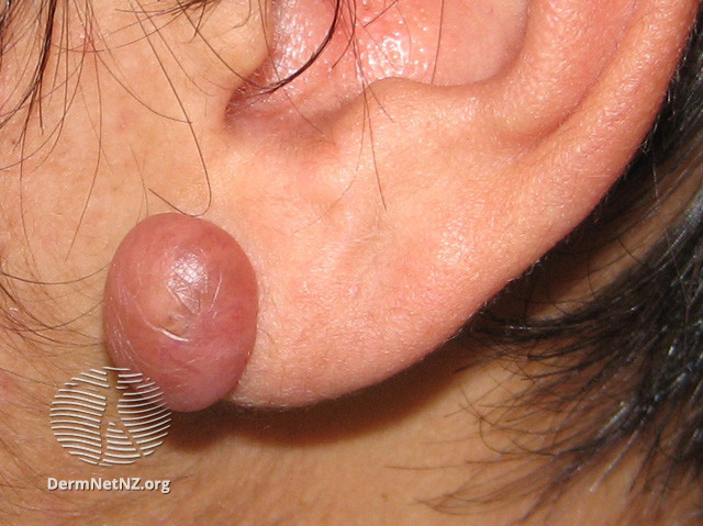
However, sometimes the process gets out of hand and exuberant which leads to hypertrophic scars (pic 1) keloids (pic 2)!

So what exactly is a scar?
Scarring is a normal part of healing that at its root, is extra collagen laid down to repair skin injury.
However, sometimes the process gets out of hand and exuberant which leads to hypertrophic scars (pic 1) keloids (pic 2)!


4/
So what's the difference between hypertrophic scars & keloids? Both have excess collagen & both can be itchy, inflamed, and bothersome.
Hypertrophic scars tend to be limited to areas of trauma, but importantly, they tend to resolve over time (months to years).
PC: @Medscape
So what's the difference between hypertrophic scars & keloids? Both have excess collagen & both can be itchy, inflamed, and bothersome.
Hypertrophic scars tend to be limited to areas of trauma, but importantly, they tend to resolve over time (months to years).
PC: @Medscape

5/
Keloids, on the other hand, tend to be larger and can continue to spread to adjacent tissues with active borders.
Histologically, keloids and hypertrophic scars have ⬆️ cellularity, vascularity, & connective tissue, but keloids have thick collagen fibers (think bubblegum).
Keloids, on the other hand, tend to be larger and can continue to spread to adjacent tissues with active borders.
Histologically, keloids and hypertrophic scars have ⬆️ cellularity, vascularity, & connective tissue, but keloids have thick collagen fibers (think bubblegum).

6/
So how do you treat them?
The mainstay of therapy for both is usually intralesional steroids. Patients often have to come back repeatedly for injections.
The best at home treatment I know of is silicone scar sheets. In fact, we used these ourselves for our daughter!
So how do you treat them?
The mainstay of therapy for both is usually intralesional steroids. Patients often have to come back repeatedly for injections.
The best at home treatment I know of is silicone scar sheets. In fact, we used these ourselves for our daughter!

7/
What if the simple stuff doesn't work?
Sometimes it's a matter of getting the actual steroid into the skin/scar. In those cases, special instruments can be tried, such as this dermajet. It literally just shoots the medication into the scar without a needle - just air...
What if the simple stuff doesn't work?
Sometimes it's a matter of getting the actual steroid into the skin/scar. In those cases, special instruments can be tried, such as this dermajet. It literally just shoots the medication into the scar without a needle - just air...

8/
And in tough cases, we'll also inject 5-fluorouracil into the scar (usually mixed with the steroids).
The 5-FU has been shown to downregulate fibroblasts, leading to decreased collagen production.
And in tough cases, we'll also inject 5-fluorouracil into the scar (usually mixed with the steroids).
The 5-FU has been shown to downregulate fibroblasts, leading to decreased collagen production.
9/
And there are a variety of surgical/physical techniques. These include laser and surgical excision.
The problem of course is any damage to the skin can more scarring, so these are often paired with medical treatment at the time of the procedure.
And there are a variety of surgical/physical techniques. These include laser and surgical excision.
The problem of course is any damage to the skin can more scarring, so these are often paired with medical treatment at the time of the procedure.
10/
People with darker skin unfortunately develop keloids and hypertrophic scars more. I don't know of any research to show why that is.
Ultimately, it's important to take a history of prior keloids/hypertrophic scars before operating on any patient.
pc:medicalnewstoday.com/articles/keloi…
People with darker skin unfortunately develop keloids and hypertrophic scars more. I don't know of any research to show why that is.
Ultimately, it's important to take a history of prior keloids/hypertrophic scars before operating on any patient.
pc:medicalnewstoday.com/articles/keloi…

11/
RECAP:
✅Scars, hypertrophic scars, & keloids are from ⬆️ collagen.
✅Keloids keep spreading, Hypertrophic scars usually stay put in areas of trauma.
✅ Lots of medical treatments available. OTC would consider silicone gel sheets!
✅ Take a keloid history before operating!
RECAP:
✅Scars, hypertrophic scars, & keloids are from ⬆️ collagen.
✅Keloids keep spreading, Hypertrophic scars usually stay put in areas of trauma.
✅ Lots of medical treatments available. OTC would consider silicone gel sheets!
✅ Take a keloid history before operating!
12/12
Hope this helps! Thanks for joining, and see you next time on another #dermtwitter #tweetorial/#medthread!
And Happy Thanksgiving!
Hope this helps! Thanks for joining, and see you next time on another #dermtwitter #tweetorial/#medthread!
And Happy Thanksgiving!
• • •
Missing some Tweet in this thread? You can try to
force a refresh


