1/
Join me for a quick #tweetorial/#medthread on:
Pearls for the #Dermatology Exam!
#MedEd #dermtwitter #medtwitter #medstudenttwitter #FOAMed
First a question - What do you think when someone asks for your help with a rash?
Join me for a quick #tweetorial/#medthread on:
Pearls for the #Dermatology Exam!
#MedEd #dermtwitter #medtwitter #medstudenttwitter #FOAMed
First a question - What do you think when someone asks for your help with a rash?
2/
Regardless how you answered, I hope to teach you something today! Let's start!
"In #dermatology, we don't do an H+P, we do a P+H."
The exam is perhaps most important. You can use it to narrow down your ddx! Then, you use your history to further work toward the right dx.
Regardless how you answered, I hope to teach you something today! Let's start!
"In #dermatology, we don't do an H+P, we do a P+H."
The exam is perhaps most important. You can use it to narrow down your ddx! Then, you use your history to further work toward the right dx.
3/
"If there's scale, there probably is epidermal involvement."
Scale usually implies action in the epidermis. This doesn't rule out anything in the dermis or subcutis, but just that the pathology includes action up top.
Check out my #tweetorial on scale
"If there's scale, there probably is epidermal involvement."
Scale usually implies action in the epidermis. This doesn't rule out anything in the dermis or subcutis, but just that the pathology includes action up top.
Check out my #tweetorial on scale
https://twitter.com/DrStevenTChen/status/1178396262833557507?s=20
4/
"If there is scale, it's probably a papule/plaque."
If you close your eyes and can palpate a difference between rash/lesion and normal skin, it's a plaque or papule (or nodule etc). And so, if you see scale, you'd probably be able to palpate that, hence: papule/plaque.
"If there is scale, it's probably a papule/plaque."
If you close your eyes and can palpate a difference between rash/lesion and normal skin, it's a plaque or papule (or nodule etc). And so, if you see scale, you'd probably be able to palpate that, hence: papule/plaque.

5/
"Distribution is the least reliable factor in the exam."
We talk about building the skin exam in layers:
Primary lesion
Secondary change
Configuration
Distribution
There's a reason "distribution" comes last, because it's the least reliable!
👀👇
"Distribution is the least reliable factor in the exam."
We talk about building the skin exam in layers:
Primary lesion
Secondary change
Configuration
Distribution
There's a reason "distribution" comes last, because it's the least reliable!
👀👇
https://twitter.com/DrStevenTChen/status/1308211387311685633?s=20
6/
"Vesicles & bullae turn into pustules over time."
You know how a transudative pleural effusion can become exudative over time (#meddermftw)? Well, same with the skin. Something that looks clear will turn purulent over time! Always be sure you're looking at the newest lesion.
"Vesicles & bullae turn into pustules over time."
You know how a transudative pleural effusion can become exudative over time (#meddermftw)? Well, same with the skin. Something that looks clear will turn purulent over time! Always be sure you're looking at the newest lesion.

7/
"Every dermatologist has been fooled by ____."
There are some relatively common diagnoses that are just tricky. I've heard this about FUNGUS (pic), and when a patient is super itchy, about SCABIES. A good dose of humility is important in dermatology (and all of medicine)!
"Every dermatologist has been fooled by ____."
There are some relatively common diagnoses that are just tricky. I've heard this about FUNGUS (pic), and when a patient is super itchy, about SCABIES. A good dose of humility is important in dermatology (and all of medicine)!

8/
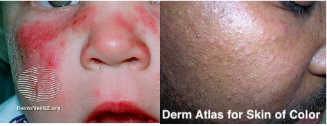
"_______ can look like anything."
On the flip side, some diagnoses can present in so many different ways, it's important to always consider them. I specifically hear this about syphilis and sarcoid (pic)!
"_______ can look like anything."
On the flip side, some diagnoses can present in so many different ways, it's important to always consider them. I specifically hear this about syphilis and sarcoid (pic)!

9/
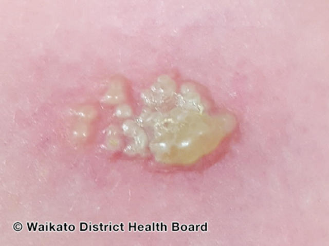
"Any erosion/ulcer is herpes until proven otherwise."
Even though he doesn't want to be the herpes guy, gotta tag @MishaRosenbach. HSV can look so atypical b/c it's been there for so long or the patient is immunosuppressed. Always think about it!
👀👇
"Any erosion/ulcer is herpes until proven otherwise."
Even though he doesn't want to be the herpes guy, gotta tag @MishaRosenbach. HSV can look so atypical b/c it's been there for so long or the patient is immunosuppressed. Always think about it!
👀👇
https://twitter.com/DrStevenTChen/status/1358189472471416835?s=20
10/10
That's all I've got for now! There are a ton more, and I'd love for #dermtwitter to pile on! Hope this is helpful for some of you out there.
Go forth and describe (...and then take a history)!
That's all I've got for now! There are a ton more, and I'd love for #dermtwitter to pile on! Hope this is helpful for some of you out there.
Go forth and describe (...and then take a history)!
• • •
Missing some Tweet in this thread? You can try to
force a refresh


